Category

volume3-issue1

464 views

Editorial

By | volume3-issue1 | No Comments

699 views

Journal of the Association of Pulmonologist of Tamil Nadu. Vol. 3, Issue 1, January – April 2020

EDITORIAL

Mediastinum

Mediastinum is the space between two medial surfaces of the right and left lung. Little did we realise when we were doing anatomy in our preclinical years that this space would be our waterloo not only in practical examinations but also in clinical practice. In our clinical years Chest x-ray was the only available tool with which one can hardly make definitive diagnosis. The pre-scan era saw many pulmonologists turning into very good radiologists with their clinical input adding on to their interpretation skills. But that was all beyond that there was no fast forward because there were no dedicated thoracic surgeons or interventional pulmonologists to get a tissue out for diagnostic purpose which would pave the way for good therapeutics. Approach to mediastinum remained a dream for many pulmonologists.

The early eighties saw the advent of imaging techniques like CAT scanning, Spiral CT, High resolution CT, contrast CT and the boom in imaging methodologies have not stopped even today. Still the tissue obtaining was an issue until the interventional pulmonologists and interventional radiologists arrived on the scene. In 1970 Conventional TBNA arrived. It was a procedure through which a needle could be introduced through the airway for sampling the mediastinal nodes. The results varied widely in several centres depending on the skills of the pulmonologists and pathologists. This became an underused technique and for 20 years there was a big lull in mediastinal approach by pulmonologists. Pulmonologists were all happy with bronchoscope that was achieving most of the airway and lung parenchymal problems with surgeons chipping to do the gold standard mediastinoscopy to sample the mediastinum.

The big leap forward came in 1990s when Endobronchial Ultrasound came as a free-standing probe and in 2004 as a scope incorporated with an ultrasound at its tip. This was a big fast forward for the pulmonologists as many latched on to this technique with ease. Even people who felt that C TBNA was a blind technique started doing this to sample the mediastinum. Results were extremely good in diagnosing tuberculosis, sarcoidosis and in staging malignancies. This has become an essential tool in the hands of the pulmonologist who can diagnose and stage the malignancy in one setting. Even PET scan which came with a big bang with regards to picking up mediastinal nodes has proved to be second to EBUS TBNA as many times PET negative sub centimetre nodes are picked by EBUS TBNA as positive for malignancy.

In all I would like to conclude interventional pulmonology is an evolving field and there is no end in sight. EBUS use is now been extended to sampling endobronchial masses and intraparenchymal masses. So there are going to be exciting times for the interventional pulmonology.

Prof. Dr. Narasimhan R, MD FRCP (E & G)

Editor-in-Chief

Journal of the Association of Pulmonologist of Tamil Nadu

How to cite this article: Narasimhan.R, Editorial, JAPT 2020; 3(1):1

874 views

To Assess the Pulmonary Impairment in Treated Pulmonary Tuberculosis Patients using Spirometry

By | volume3-issue1 | No Comments

699 views

V.G. Vinod et al.: To Assess the Pulmonary Impairment in Treated Pulmonary Tuberculosis Patients using Spirometry

Original Study

To Assess the Pulmonary Impairment in Treated Pulmonary Tuberculosis Patients using Spirometry

V.G.Vinod1, S.M. Soumya2, M. Jayashree2, S. Darshini2, T. Dhanasekar3

1Assistant Professor, Sri Ramachandra Institute of Higher Education and Research
2Respiratory Therapist, Sri Ramachandra Institute of Higher Education and Research
3Professor, Sri Ramachandra Institute of Higher Education and Research

Abstract

Background: Despite proper completion of treatment, pulmonary TB (PTB) patients experience pulmonary dysfunction which can cause significant impact in their quality of life. This study aimed to assess the lung function impairment in patients treated for PTB using spirometry. Methods: A cross-sectional study was conducted in Pulmonary Medicine Department Out-patient department in Sri Ramachandra Institute of Higher Education and Research. Patients aged 15 and above who were treated for pulmonary tuberculosis were included in the study. Demographic data were collected. All participants underwent spirometric measurements. Airflow obstruction was defined as apost- bronchodilation FEV1/FVC < 70% with FVC > 80%, restrictive defects as an FEV1/FVC ratio of ≥ 70% with an FVC < 80% predicted, and mixed defects as FVC of <80% predicted and an FEV1/FVC ratio of <70%. Lung function impairment was defined by the presence of at least one of these three abnormalities. Results: Of a total of 20 participants included in the study, 10 (50%) were male. The mean age of participants was 52 years. The mean BMI of the participants was 21.70. Spirometry showed ventilatory impairment in 95%of which obstructive pattern was the most common ventilatory impairment noted.Conclusion: Post-tuberculous pulmonary function impairment is very highly prevalent. Predominant impairment found is obstructive ventilator defect. Early and prompt diagnosis of such pulmonary dysfunction can improve their quality of life with pharmacotherapy and rehabilitation.

Corresponding Author: Dr. V.G.Vinod Assistant Professor Department of pulmonology Sri Ramachandra Institute of Higher Education and Research Porur, Chennai Tamil nadu, India. Email: v.g.vinod14@gmail.com@gmail.com

Background

Pulmonary Tuberculosis, the global epidemic, is caused by the acid fast bacilli Mycobacterium tuberculosis. It is still one of the leading infectious diseases in the world (1). Tuberculosis is one of India’s major public

health problems. The incidence of tuberculosis in India according to a recent report is 204/100,000 population (2).In India, RNTCP has been making effective strategies for the control and elimination of TB. But still pulmonary tuberculosis is still prevalent. Not only active disease, but also post infectious sequelae is very common causing heavy burden on the health

How to cite this article: V.G.Vinod, S.M. Soumya, M. Jayashree, S. Darshini, T. Dhanasekar, To Assess the Pulmonary Impairment in Treated Pulmonary Tuberculosis Patients using Spirometry, JAPT 2020; 3(1):3-6

system. Despite completion of treatment and microbiological cure, myriad defects are noted on the anatomy and pathophysiology in pulmonary tuberculosis patients. Sequelae can involve any component of pulmonary architecture ranging from airway to mediastinum. After treatment of TB, the lung is left with residual fibrosis, scarring, cavitation and distortion of lung architecture leading to volume loss, and bronchiectasis (3). Depending on the site and extent of involvement, patients can have a wide variety of symptoms ranging from minimal symptoms to severe breathlessness (4). An attempt was made to assess the prevalence of impairment of respiratory function in treated pulmonary tuberculosis patients.

Methods

It is a prospective observational study carried out in Pulmonary Medicine Department from December 2019 to January 2020, Sri Ramachandra Institute of Higher Education and Research [SRIHER]. A total of 20 cases were included in the study.

Inclusion criteria

Patients who were of aged more than 18 years and has completed Pulmonary tuberculosis treatment for 6 months and declared cured.

Exclusion criteria

Patients with Irregular Tuberculosis treatment history, Acute COPD exacerbations, Cardiovascular diseases, Active infections, Haemoptysis, Hypertension, Recent abdominal surgery, Recent eye surgeries, Recent MI, Extra pulmonary TB cases, Multidrug resistant TB cases, Cases unable to cooperate during spirometry procedure or does not want to enroll in the study, Cases who failed to fulfil acceptability and reproducibility criteria of spirometry were excluded from the study, Defaulted cases during treatment.

Procedure

Patients who are diagnosed with tuberculosis sequelae by pulmonologist in the outpatient department were taken for the study. The patient’s medical history was checked for relevant details and data procured. Patient’s height and weight were checked. Inclusion, Exclusion criteria were applied and if the patient qualified for the study He/She was taken up for the study and was explained about spirometry.
Subsequently, spirometric evaluation was done and meets American Thoracic Society and European Respiratory Society standards (ATS and ERS), before and 15 minutes after administration of 400 microgram salbutamol via metered-dose inhaler. All patients were instructed not to use any bronchodilator on the preceding night and on the day of procedure. Spirometric procedure was carried out as per ATS/ERS task force recommendation for standardization of lung function testing. FVC, FEV1, and FEV1/FVC ratio values for case patients were compared with gender-specific and race-specific adult predicted normative population values and the control group.
Interpretive algorithms were used in determining restrictive or obstructive patterns and spirometry results were analyzed and categorized in four groups as follows: (5, 6, 7, 8)
1. Normal – FEV1/FVC ratio of >70% and an FVC of >80% predicted
2. Obstructive – airway obstruction was defined as an FEV1/FVC ratio of <70% and an FVC of >80% predicted
3. Mixed-combined defects were FVC of <80% predicted and an FEV1/FVC ratio of <70% 4. Restrictive-restrictive defects as an FEV1/FVC ratio of >70% with an FVC of <80% predicted.

Results

Of the 20 cases, there were 10 males (50%) and 10 females (50%). Of those, 15% (3/20) were between age group of 30 – 40 years; 20% (4/20)
were between 40-50 years; 35% (7/20) were between age group of 50-60 years and 30% (6/20) were between 60-70 years (Table 1).


Table 1: Age distribution of post-tuberculosis cases

In the study group, prevalence of smoking was 45% (9/20). 90% (9/10) males were smokers but none of the female had smoking history (Table 2).


Table 2: Smoking status of post-tuberculosis cases

Of the 20 participants, 15% (3/20) were underweight, 70% (14/20) were normal weight and 15% (3/20) were overweight (Table 3).


Table 3: BMI status of post-tuberculosis cases

In spirometry assessment of the study group, abnormal lung function is documented in 95% (19/20) cases. The obstructive ventilatory pattern was the predominant pattern which was documented in 50% (10/20) cases; Restrictive ventilatory pattern was documented in 25% (5/20) cases; Mixed ventilatory pattern was documented in 20% (4/20) cases and normal spirometry was documented in 5% (1/20) cases (Table 4).


Table 4: Spirometry assessment of post-tuberculosis cases

Discussion

The mean age of the study was 52 years. This was comparable to the study done by Mozaffari et al (9) who had 48.31 as the mean age in their study. Ngahane et al (10) had mean age of 34.2 years which could be due to variations in study population.
Males and females were of equal numbers in the current study which was closely resembling the study population of Ngahane et al. who had 54.3% male and 45.7 % females. But Mozaffari et al. had around 1/3rd males in their study population which could be due to difference in incidences of the disease in different geographical locations.
Mean BMI of the present study was 21.70. It was comparable to study done by Ngahane et al (10) who had 23.4 as the mean BMI in their study. Akshay N.Gupte et al (11) had mean BMI of 32 which is high comparing to our study probably due to the socioeconomic differences between the study group.
45% of our study population were smokers which is similar to the study done by Ramos et al (12) who had 46% of their study population as smokers (former smokers + current smokers). Ngahane et al had 10% smokers in their study group which is low comparing to our study – variation is likely due to cultural characteristics differences between the study population.
Obstructive ventilatory pattern is the common pulmonary function abnormality noted in our study population which is similar to the studies done by Shital patel et al (13) and sailaja et al (14) who had obstructive pattern as the predominant abnormality in their study too. But Ngahane et al had restrictive pattern as the predominant pulmonary function defect in their study which could be due to difference in race between the study populations.

Conclusion

The present study reiterates that tuberculosis sequelae is highly prevalent , most of which causing an obstructive pattern, although restrictive and mixed patterns are also
present. So pulmonary tuberculosis patients have to be followed up even after the completion of treatment for early diagnosis of lung impairment due to pulmonary tuberculosis sequelae. Such patients have to be evaluated and managed with bronchodilators and pulmonary rehabilitation.

Reference

1. Hershkovitz I, et al., Tuberculosis origin: The Neolithic scenario, Tuberculosis (2015), http://dx.doi.org/10.1016/ j.tube.2015.02.021
2. World Health Organization. Global Tuberculosis Report 2018. Geneva, Switzerland: World Health Organization; 2018
3. Dheda K, Booth H, Huggett JF, Johnson MA, Zumla A, Rook GA. Lung remodeling in pulmonary tuberculosis. J Infect Dis. 2005;192(7):1201-1209. doi:10.1086/444545
4. Musafiri S, Dusabejambo V, Munganyinka BC, Manzi O, Kalisa L and Rutayisire PC. The Aftermath of Pulmonary Tuberculosis: Predictors of Severe Pulmonary Sequelae and Quality of Life of Patients Visiting a Tertiary Level of Care in Rwanda, East Africa. Austin J Pulm Respir Med 2015; 2(2): 1027. ISSN:2381-9022
5. Crapo RO, Morris AH, Gardner RM. Reference spirometric values using techniques and equipment that meet ATS recommendations. Am Rev Respir Dis 1981;123:659-64.
6. Miller MR, Hankinson J, Brusasco V, Burgos F, Casaburi R, Coates A, et al. Standardisation of spirometry. Eur Respir J 2005;26:319-38.
7.Evaluation of impairment/disability secondary to respiratory disorders. American Thoracic Society. Am Rev Respir Dis 1986;133:1205-9.
8.Enright PI, Hyatt RE. Office Spirometry: A Practical Guide to Selection and Use of Spirometers. Philadelphia, PA: Lea & Febiger; 1987.
9. European Respiratory Journal 2018 52: Suppl. 62, PA2747.
10. Mbatchou Ngahane BH, Nouyeb J, Nganda Motto M Yacouba MN, Wandji A, Endalle M, Afane Ze E, Post- tuberculous lung function impairment in a tuberculosi reference clinic in Cameroon, Respiratory Medicine (2016), doi: 10.1016/j.rmed.2016.03.007.
11.Gupte, Akshay N et al. “Assessment of lung function in successfully treated tuberculosis reveals high burden of ventilatory defects and COPD.” PloS one vol. 14,5 e0217289. 23 May. 2019, doi:10.1371/journal.pone.0217289
12. Ramos LM, Sulmonett N, Ferreira CS, Henriques JF, de Miranda SS. Functional profile of patients with tuberculosis sequelae in a university hospital. J Bras Pneumol. 2006;32(1):43-47. doi:10.1590/s1806- 37132006000100010
13. Patil S et al. Pulmonary functions’ assessment in post- tuberculosis cases by spirometry: Obstructive pattern is predominant and needs cautious evaluation in all treated cases irrespective of symptoms. Int J Mycobacteriol 2018;7:128-33
14. Sailaja K, Nagasreedhar Rao H. Study of pulmonary function impairment by spirometry in post pulmonary tuberculosis. J Evolution Med Dent Sci. 2015;4(42):7365- 70. http://dx.doi.org/10.14260/jemds/2015/1068

453 views

Early Predictors of Success of Non-Invasive Ventilation in Acute Hypercapnic Respiratory Failure

By | volume3-issue1 | No Comments

699 views

Umesh G et al.: Early Predictors of Success of Non-Invasive Ventilation in Acute Hypercapnic Respiratory Failure

Original Study

Early Predictors of Success of Non-Invasive Ventilation in Acute Hypercapnic Respiratory Failure

Umesh G1, Winnie Elizabeth Jose2, Sundararajan L3

1Pulmonologist, Primary Health Centre Cheruthana, Alazpuzha District, Kerala.
2Senior resident, Alapuzha Government Medical College, Kerala
3Senior Consultant Pulmonologist, Department of Respiratory Medicine, Apollo Hospitals, Chennai, Tamilnadu.

Abstract

Introduction: Non-invasive ventilation (NIV) has emerged as a significant advancement in the management of acute hypercapnic respiratory failure. Non-invasive positive pressure ventilation (NIV) using face mask or nasal mask which works by providing pressure support that gives ventilatory
assistance during inspiration, allows respiratory muscles to work less, increases the volume inspired per minute and improves arterial blood gas (ABG) levels. Its use has become more common as its benefits are increasingly recognized. However, the frequency of success of NIV in patients with acute respiratory failure varies amongst studies. In some patients, with acute hypercapnic respiratory failure, NIV is inadequate and invasive ventilation is required for management of respiratory failure. Failure of initial trial of NIPPV can lead to a delay in endotracheal intubation thus causing an increase in morbidity and mortality. Thus, determination of early predictors of success of NIPPV is important to identify the patients who are likely to benefit from NIPPV.
Aim of The Study: To identify the early predictors of success of non-invasive positive pressure ventilation in patients admitted with acute hypercapnic respiratory failure.
Materials and Methods: Patients with acute hypercapnic respiratory failure requiring ventilation therapy with respiratory rate [RR] of > 23 breaths per minutes, PaCO2 > 45 mm Hg and arterial pH < 7.35 (despite maximum medical treatment on controlled oxygen therapy) were included in the study. Baseline parameters such as heart rate (HR), respiratory rate (RR), oxygen saturation (SPO2) and Arterial blood gas [ABG] were collected and the same had been re-evaluated at 2, 24 hours and before discharge after initiation of NIV. Change in these parameters, need for intubation and survival to discharge were analyzed.
Results: Total number of patients included was 50; 54% were males and 46% were females. Mean age was 65.2 ± 10.2 yrs. Overall survival to discharge with NIV management was 74%. Intubation rate was 34%. No death occurred during NIV. Average length of stay in NIV success group was 7.32 ± 2.25 days.

How to cite this article: Umesh G, Winnie Elizabeth Jose, Sundararajan L, Early Predictors of Success of Non-Invasive Ventilation in Acute Hypercapnic Respiratory Failure, JAPT 2020; 3(1):7-15

Statistical significance observed in variables such as pH , PaCO2, SPO2, HR and RR at baseline and statistical improvement observed in pH , HR, RR at 2 hours after initiation of NIV. ROC curve analysis showed a value below baseline pH of 7.14, SPO2 below 79% and 2 hr pH of 7.32, had higher chance of NIV failure. Conclusion: Early predictors such as degree of acidosis, PaO2, PaCO2, HR, RR, SPO2 have significant role in identifying patients who were benefitted by NIV thus preventing the delay in initiation of invasive ventilation.
Keywords: Acute hypercapnic respiratory failure, NIV, intubation rate, early predictors of success

Corresponding Author: Dr.Umesh. G, Pulmonologist, Primary Health Centre Cheruthana, Alazpuzha District, Kerala. Email:drumeshchest@gmail.com

Introduction

Mechanical ventilation (MV) is a form of life support measure for patients with acute respiratory failure. The primary aim is to achieve adequate alveolar ventilation along with improved oxygen exchange by providing mechanical breaths1,2. Mechanical ventilation can be delivered invasively or non-invasively. Invasive mechanical ventilation is associated with a host of attendant complications, like ventilator-associated pneumonia, barotrauma, inability to speak, swallow or eat, and laryngeal trauma while the tube is in place or after its removal.2,3

Non-invasive ventilation (NIV) refers to the administration of ventilator support without using an invasive artificial airway (end otracheal tube or tracheostomy tube). The use of non-invasive ventilation has markedly increased over the past two decades and NIV has now become an integral tool in the management of both acute and chronic respiratory failure in both home setting and in the critical care unit. Its use in acute respiratory failure is well accepted and widespread1.
Non-invasive positive pressure ventilation (NIPPV) works by providing pressure support that gives ventilatory assistance during inspiration, allows respiratory muscles to work less, increases the volume inspired per minute and improves arterial blood gas (ABG) levels4,5. Nowadays only positive pressure ventilation is in use worldwide so NIV is synonymous with NIPPV.
In some patients, with acute hypercapnic respiratory failure, NIV is inadequate and invasive ventilation is required for management of respiratory failure. The failure rates in such patients range from 7% to 50% in different studies. Failure of initial trial of NIV can lead to a delay in invasive mechanical ventilation thus causing an increase in morbidity and mortality. Thus, determination of early predictors of success of NIV is important to identify the patients who are likely to benefit from NIV6, 7, 8. Patients responding to NIV will be continued with NIV.
This study was designed to primarily assess the safety and efficacy of NIV in acute hypercapnic respiratory failure in a tertiary care hospital and additionally to identify prognostic variables for survival, death and endotracheal intubation.

Methodology

Patients presenting with acute hypercapnic respiratory failure, not improving initially with optimal medical therapy and controlled oxygen therapy and meeting criteria for non-invasive ventilation were included during the study period [December 2015-November 2016]. Hospital ethical committee clearance was obtained before the study was started. Informed consent was taken from all participants in the study. All patients were managed in the intensive care unit. A portable non-invasive ventilator was used in the spontaneous mode or through ICU ventilator using or onasal mask as decided by attending critical care physician / Intensivist depending on patient’s clinical condition. The inspiratory pressure support was initially set at 15 cm of water. Expiratory pressure was set at 3 cm of water. The pressure was gradually increased to reach the optimum level for each patient. Oxygen was administered through the NIV interface to maintain oxygen saturation (SpO2) between 88% to 92%. The
patient’s electrocardiogram (ECG), SpO2, blood pressure (BP), and respiratory rate (RR) were continuously monitored. Standard medical treatment including inhalational bronchodilators, intravenous corticosteroids, xanthene and whenever appropriate antibiotics, diuretics, or vasopressors were given in addition to NIV. Clinical history was obtained from the patients or from family members at the time of admission. A thorough clinical examination was carried out. RR, blood pressure, oxygen saturation (SpO2), and ECG were continuously monitored. Arterial blood gas analysis was done before the patient was put on NIV (i.e. baseline) and serially monitored at 2 hours, 24 hours and at the time of discharge.

The mask was examined for leak, skin abrasion, and patient satisfaction. If satisfactory degree of patient comfort, ventilation and oxygenation were not achieved, ABG was repeated. Patients were put on invasive MV when there was any deterioration. On the first day, patients were put on NIV for 24 hours and on the following days depending upon the acidosis and clinical condition, the NIV duration was gradually decreased if there was improvement, ABG was also taken at the time of discharge. Variables at different time in both were subjected to statistical analysis. Data with P values < 0.05 were considered statistically significant. Long term oxygen or NIV was prescribed at the time of discharge if needed.

Definition of Success and Failure

Success was defined as the achievement of clinical and functional condition stable enough to allow patient discharge without invasive mechanical ventilation either by endotracheal intubation or tracheostomy.

Patients who were put on invasive mechanical ventilation or dead were to decided to have failed NIV.

Intubation Criteria

After initiation of NIV, invasive mechanical ventilation was considered if;

  • Deterioration in patient’s condition
  • Failure to improve or deterioration in arterial blood gas tensions
  • Development of new symptoms or complications such as pneumothorax, sputum retention, nasal bridge erosion
  • Intolerance or failure of coordination with the ventilator
  • Failure to alleviate symptoms
  • Deteriorating conscious level
  • Patient and care-taker wish to withdraw treatment

A management plan of what to do if NIV fails was made early and finally the decision to progress to intubation was made by an experienced clinician in consultation with ICU staff.

Results & Observations

Total of fifty patients were included in the study, thirty-seven (74%) patients admitted with acute hypercapnic respiratory failure improved clinically with initial trial of NIV and continued with NIV and thirteen (26%) patients failed the initial trial of NIV and got intubated. Out of 13 in failure group, 4 patients got intubated after 24 hours. [chart 1]. Total of fifty patients were included in this study of which 27(54%) were males and 23(46%) were females. The following pie diagram represents percentage gender distribution. The mean age of total study population was 65.2 ± 10.2[range: 34-85]. The mean age was 63.83± years in success group and 69.07 ±years in failure  group and there was no stasticially significance difference between the two groups.

In our study co-morbidities associated with acute hypercapnic respiratory failure were COPD (27), OSA (6), BA (1), DPLD (5) most ILD with UIP pattern cardiogenic pulmonary oedema (1), pneumonia (1) and other in combination such as COPD and OSA (3), COPD and Pneumonia (5), BA and OSA (1)  [chart:2.].  The co-morbidities were cardiac disease, diabetes mellitus,  hypertension, hypothyroidism, chronic kidney  disease and it was found that these co-morbidities were almost equally among both groups.

Serial Monitoring of Study Variables:

The changes observed the mean of variables such as pH, PaCO2, PaO2, SPO2, HR, RR in the success group at different time showed linear improvement towards normal value more than that of failure group which showed slow improvement even after 24 hours. The error graphs [grap1, 2 & 3] showing changes in the significant values (pH, at baseline and at 2 hrs of NIV between two groups.

Receiver operating characteristics [ROC]curve analysis was done to find the cut off values of significant predictor variables. Baseline values of pH below 7.14 (sensitivity of 100% and specificity of 64%) and SPO2 less than 79% (sensitivity of 86 % and specificity of 72 %) had more chance of NIV failure and lack of improvement of PH even after 2 hours more than 7.32 (sensitivity of 62% and specificity of 100%) had higher chance of NIV failure.

Discussion

NIV is a better alternative for invasive mechanical ventilation in patients presenting  with acute hypercapnic respiratory failure, who  are in need of ventilator support to improve gas exchange in our study NIV was successful in 74% of patient which is comprable to other similar studies published by  Bhattacharya et  al8  and Salahuddin et al6. We selected patients  according to inclusion and exclusion criteria as per recognised NIV guidelines.

Delay in starting invasive mechanical  ventilation to whom it would be benefitted after  initiation of NIV can increase mortality and  morbidity. So, variables predicting the outcome of NIV will be useful. Several studies  so far published6,8,5,7,6,4 observed that ABG values, respiratory rate and heart rate found at baseline have significant role in early prediction of NIV success.
In our study we observed baseline higherpH, oxygen saturation (SPO2) and lower respiratory rate, heart rate significantly (p value
< 0.05) predicted the success of NIV. Other variable such as PCO2, bicarbonate, PaO2 did not show any significance difference unlike in study done by Bhattacharya et al.8 Singh VK et al. observed high baseline heart rate in failure group5. Conti et al. 64 and Ambrosino et al. 65 in
their study showed high baseline pH in success group.
After 2 hour, statistically significant improvement was observed in pH, heart rate and respiratory rate in success group compared to failure group but not in other variables. The same has been observed in other studies also5,8.

On ROC curve analysis, pH less than 7.14 at baseline, SPO2 less than 79% and no improvement in pH after 2 hrs more than 7.32 had higher chances of NIV failure. Salahuddin et al observed higher risk of NIV failure if baseline pH less than 7.30, serum bicarbonate less than 35 meq / L.

In success group it was observed that there was a gradual improvement in ABG values, HR and RR towards normal even after 24 hour and at the time of discharge.

Salahuddin et al8. in their study observed that younger age has more success rate, however in our study, we could not find any statistical significance in the term of age factor.

Various studies have proved the success rate of NIV in COPD patients but very few studies have published on the outcome of NIV in alternative aetiologies of acute hypercapnic respiratory failure. Respiratory failure associated with COPD had comparatively better success rate when compared with others6. Our study had heterogenous group of diseases including, COPD, OSA, Bronchial asthma, pneumonia, interstitial lung disease and cardiogenic pulmonary oedema. We could not analyse individual diseases since majority of the study population were COPD patients.

Pneumonia was an independent predictor of outcome, observed by various studies6,65,66. Angela Maria Grazia Pacilli et al.66 observed pneumonia as an independent predictor of failure with 45%. Similarly, in our study pneumonia was associated with more chance of NIV failure (p value 0.015, odds ratio 3.36).

In our the study we found multiple co-morbidities such as diabetes mellitus, hypertension, cardiac disease, hypothyroidism and chronic kidney disease in combination or individually distributed almost equally among both groups. Patil SP et al67 showed an increased chance of hospital mortality in the presence of more co morbidities. But in another study, Seneff et al68 demonstrated that, there was no significance in the number of co morbidities and outcome or NIV.

The average length of stay in success group was 7.32 ± 2.25 days which was less than other studies8.
Thirteen patients failed the initial trial of NIV and got intubated. No deaths were observed during NIV trial. Out of 13, 9 patients got intubated before 24 hours and 4 patients after 24 hours. Reasons for intubation were worsening symptoms with severe acidosis even after initial NIV trial, cardiac and respiratory arrest, patient asynchrony, increased sputum production and low GCS.

Error bar showing the difference in PH across time points

Conclusion

Data available at the time of initiation NIV and after 2 hrs can predict the likelihood of success or failure with a reasonable degree of precision. The severity of acidosis at baseline with other variables such as heart rate, respiratory rate, PaCO2 were indetifies as an important predictor; although NIV is less likely to be effective when patients are more acidotic. This should not preclude a trial of NIV as the mode of ventilatory support of first choice because the benefits of NIV compared with intubation and mechanical ventilation are greater. The tolerance of NIV and the change in arterial blood gas tensions, particularly pH, PCO2, respiratory rate and heart rate in the early hours are reasonable predictors of the  subsequent outcome. Patient should be co- operative, have the ability to protect airway. Appropriate pressure settings, good patient machine synchronisation and clinical judgement by the attending physician and medical team are crucial in alternating the success of NIV.

Reference

1. Bello, Giuseppe, Gennaro De Pascale, and Massimo Antonelli. “Non-invasive Ventilation”. Clinics in Chest Medicine 37.4 (2016): 711-721.
2. Nava, Stefano and Nicholas Hill. “Non-Invasive Ventilation in Acute Respiratory Failure”. The Lancet 374.9685 (2009): 250-259.
3. Hill, Nicholas S. et al. “Non-invasive Ventilation in Acute Respiratory Failure”. Critical Care Medicine 35.10 (2007): 2402-2407.
4. Sharma, RM, A Handa, and R Chaturvedi. “Invasive toNon-invasive Ventilation in Chronic Obstructive Pulmonary Disease”. Medical Journal Armed Forces India 59.3 (2003): 264-265.
5. Singh VK, Khanna P, Rao BK, Sharma SC, Gupta R.” Outcome prediction for non-invasive positive pressure ventilation in acute respiratory failure”. JAPI 2006; 54:361–365.
6. Salahuddin, N, M Naeem, and S Khan. “Physiological Variables Predictive Of Survival In Patients With Acute Type II Respiratory Failure On Non-invasive Ventilation”. Critical Care11. Suppl 2 (2007): P168. Web.
7. Antonello Nicolini, Ines Maria Grazia Piroddi, et al. “Predictors of Non-Invasive Ventilation Failure in Severe Respiratory Failure Due to Community Acquired Pneumonia”. Tanaffos. 2014; 13(4): 20–28.
8. Bhattacharyya, D et al. “Early Predictors Of Success Of Non-Invasive Positive Pressure Ventilation In Hypercapnic Respiratory Failure”. Medical Journal Armed Forces India 67.4 (2011): 315-319. Web.
9. Drinker, Philip and Louis A. Shaw. “An apparatus for the prolonged administration of artificial respiration”. Journal of Clinical Investigation 7.2 (1929): 229-247. Web.
10. Woolam, C. H. M. “The Development Of Apparatus For Intermittent Negative Pressure Respiration (2) 1919?1976, With Special Reference To The Development And Uses Of Cuirass Respirators”. Anaesthesia 31.5 (1976): 666-685. Web.
11. Mehta, Sangeeta and Nicholas s. hill. “Non-invasive Ventilation”. American Journal of Respiratory and Critical Care Medicine 163.2 (2001): 540-577. Web.
12. Barach AL, Martin J, Eckman M. “Positive pressure respiration and its application to the treatment of acute pulmonary oedema”. Annals of Internal Medicine 12.6 (1938): 754. Web.
13. “Intermittent Positive Pressure Breathing Therapy Of Chronic Obstructive Pulmonary Disease”. Annals of Internal Medicine 99.5 (1983): 612. Web.
14. Klein G, Ruhle KH, Matthys H. “Long-term oxygen therapy vs. IPPB therapy in patients with COPD and respiratory insufficiency: survival and pulmonary hemodynamics”. Eur J Respir Dis Suppl. 1986;146: 409-415.
15. Bach, John R. et al. “Mouth Intermittent Positive Pressure Ventilation In The Management Of Postpolio Respiratory Insufficiency”. Chest 91.6 (1987): 859-864. Web.
16. Bach, John R. et al. “Management Of End Stage Respiratory Failure In Duchenne Muscular Dystrophy”. Muscle & Nerve 10.2 (1987): 177-182. Web.
17. Bach, John R. and Augusta S. Alba. “Management Of Chronic Alveolar Hypoventilation By Nasal Ventilation”. Chest 97.1 (1990): 52-57. Web.
18. Sullivan, Colin E. and Elizabeth R. Ellis. “Nocturnal Positive Pressure Ventilation Via A Nasal Mask”. American Review of Respiratory Disease 136.3 (1987): 791- 792. Web.
19. Ellis ER, Bye PT, Bruderer JW, Sullivan CE. Treatment of respiratory failure during sleep in patients with neuromuscular disease. Positive-pressure ventilation through a nose mask. American Review of Respiratory Disease. 1987 Jan;135(1):148-52.
20. De Paoli AG, Davis PG, Faber B, et al. “Devices and pressure sources for administration of nasal continuous positive airway pressure (NCPAP) in preterm neonates”. Cochran Database Syst Rev. 2008;1:CD002977.
21. Liesching, Timothy, Henry Kwok, and Nicholas S. Hill. “Acute Applications Of Non-invasive Positive Pressure Ventilation*”. Chest 124.2 (2003): 699-713. Web.
22. Ambrosino N, Vagheggini G. “Non-invasive positive pressure ventilation in the acute care setting: where are we?” Eur Respir J (2008) 31:874-886
23. Keenan, Sean P. et al. “Does Non-invasive Positive Pressure Ventilation Improve Outcome In Acute Hypoxemic Respiratory Failure? A Systematic Review”. Critical Care Medicine 32.12 (2004): 2516-2523. Web.
24. Crane SD, Elliot MW, Gilligan P, et al; “ Noninvasive ventilation in cardiogenic pulmonary edema: A multicenter randomized trial”; Am Journal Respir Crit Care Med 2003; 168: 1432-1437
25. Crane, S D. “Randomised Controlled Comparison Of Continuous Positive Airways Pressure, Bilevel Non- Invasive Ventilation, And Standard Treatment In Emergency Department Patients With Acute Cardiogenic Pulmonary Oedema”. Emergency Medicine Journal 21.2 (2004): 155-161. Web.
26. Mehta, Sangeeta et al. “Randomized, Prospective Trial Of Bilevel Versus Continuous Positive Airway Pressure In Acute Pulmonary Edema”. Critical Care Medicine 25.4
(1997): 620-628. Web.
27. Jolliet, Philippe et al. “Non-Invasive Pressure Support Ventilation In Severe Community-Acquired Pneumonia”. Intensive Care Medicine 27.5 (2001): 812- 821. Web.
28. CONFALONIERI, MARCO et al. “Acute Respiratory Failure In Patients With Severe Community-Acquired Pneumonia”. American Journal of Respiratory and Critical Care Medicine 160.5 (1999): 1585-1591. Web.
29. Ferrer, Miquel et al. “Noninvasive Ventilation In Severe Hypoxemic Respiratory Failure”. American Journal of Respiratory and Critical Care Medicine 168.12 (2003): 1438-1444. Web.
30. Honrubia, Teresa et al. “Noninvasive Vs Conventional Mechanical Ventilation In Acute Respiratory Failure”. Chest 128.6 (2005): 3916-3924. Web.
31. Antonelli, Massimo et al. “A Multiple-Center Survey On The Use In Clinical Practice Of Noninvasive Ventilation As A First-Line Intervention For Acute Respiratory Distress Syndrome*”. Critical Care Medicine 35.1 (2007): 18-25. Web.
32. Nourdine, K. et al. “Does Noninvasive Ventilation Reduce The ICU Nosocomial Infection Risk? A Prospective Clinical Survey”. Intensive Care Medicine 25.6 (1999): 567-573. Web.
33. Plant, PK, JL Owen, and MW Elliott. “Early Use Of Non-Invasive Ventilation For Acute Exacerbations Of Chronic Obstructive Pulmonary Disease On General Respiratory Wards: A Multicentre Randomised Controlled Trial”. The Lancet 355.9219 (2000): 1931-1935. Web.
34. Brochard, Laurent et al. “Noninvasive Ventilation For Acute Exacerbations Of Chronic Obstructive Pulmonary Disease”. New England Journal of Medicine 333.13 (1995): 817-822. Web.
35. Lightowler, J. V. “Non-Invasive Positive Pressure Ventilation To Treat Respiratory Failure Resulting From Exacerbations Of Chronic Obstructive Pulmonary Disease: Cochrane Systematic Review And Meta- Analysis”. BMJ 326.7382 (2003): 185-185. Web.
36. Keenan, Sean P. “Which Patients With Acute Exacerbation Of Chronic Obstructive Pulmonary Disease Benefit From Noninvasive Positive-Pressure Ventilation?”. Annals of Internal Medicine 138.11 (2003): 861. Web.
37. Vignaux, Laurence, Didier Tassaux, and Philippe Jolliet. “Performance Of Noninvasive Ventilation Modes On ICU Ventilators During Pressure Support: A Bench Model Study”. Intensive Care Medicine 33.8 (2007): 1444-1451. Web.
38. Ferreira, Juliana C., Daniel W. Chipman, and Robert M. Kacmarek. “Trigger Performance Of Mid-Level ICU Mechanical Ventilators During Assisted Ventilation: A Bench Study”. Intensive Care Medicine 34.9 (2008): 1669-1675. Web.
39. Pennock, Bernard E., Linda Crawshaw, and Peter D. Kaplan. “Noninvasive Nasal Mask Ventilation For Acute Respiratory Failure”. Chest 105.2 (1994): 441-444. Web.
40. Joris, Jean L. et al. “Effect Of Bi-Level Positive Airway Pressure (Bipap) Nasal Ventilation On The Postoperative Pulmonary Restrictive Syndrome In Obese Patients Undergoing Gastroplasty”. Chest 111.3 (1997): 665-670. Web
41. Robert M. Kacmarek; “Non-invasive Positive Pressure Ventilation: The little things do make the difference!” Respiratory Care 2003;48: 919-921
42. Boitano LJ. Management of airway clearance in neuromuscular disease. Respir Care 2006; 51:913.
43. Dean R Hess; “Noninvasive Ventilation in Neuromuscular Disease: Equipment and Application”. Respir Care 2006;51(8):896–911.
44. Navalesi, Paolo et al. “Physiologic Evaluation Of Noninvasive Mechanical Ventilation Delivered With Three Types Of Masks In Patients With Chronic Hypercapnic Respiratory Failure”. Critical Care Medicine 28.6 (2000): 1785-1790. Web.
45. Lemyze, Malcolm et al. “Rescue Therapy By Switching To Total Face Mask After Failure Of Face Mask- Delivered Noninvasive Ventilation In Do-Not-Intubate Patients In Acute Respiratory Failure*”. Critical Care Medicine 41.2 (2013): 481-488. Web.
46. Girault, Christophe et al. “Interface Strategy During Noninvasive Positive Pressure Ventilation For Hypercapnic Acute Respiratory Failure*”. Critical Care Medicine 37.1 (2009): 124-131. Web.
47. “International Consensus Conferences In Intensive Care Medicine: Noninvasive Positive Pressure Ventilation In Acute Respiratory Failure”. American Journal of Respiratory and Critical Care Medicine 163.1 (2001): 283-291. Web.
48. Goodacre, Steve et al. “Prehospital Noninvasive Ventilation For Acute Respiratory Failure: Systematic Review, Network Meta-Analysis, And Individual Patient Data Meta-Analysis”. Academic Emergency Medicine 21.9 (2014): 960-970. Web.
49. Nava, Stefano, Paolo Navalesi, and Giorgio Conti. “Time Of Non-Invasive Ventilation”. Intensive Care Medicine 32.3 (2006): 361-370. Web.
50. Nava S, Navalesi P, Conti G. “Time of non-invasive ventilation”. Intensive Care Med 2006; 32:361.
51. Collaborative Research Group of Noninvasive Mechanical Ventilation for Chronic Obstructive Pulmonary Disease. Early use of non-invasive positive pressure ventilation for acute exacerbations of chronic obstructive pulmonary disease: a multicentre randomized controlled trial. Chin Med J (Engl) 2005; 118:2034.
52. Ozsancak Ugurlu, Aylin et al. “Use And Outcomes Of Noninvasive Positive Pressure Ventilation In Acute Care Hospitals In Massachusetts”. Chest 145.5 (2014): 964-971. Web.
53. Liesching, Timothy, Henry Kwok, and Nicholas S. Hill. “Acute Applications Of Noninvasive Positive Pressure Ventilation*”. Chest 124.2 (2003): 699-713. Web.
54. Kacmarek, RM. “Characteristics of pressure-targeted ventilators used for noninvasive positive pressure ventilation”. Respir Care 1997; 42:380.
55. Ferguson, G T and M Gilmartin. “CO2 Rebreathing During Bipap Ventilatory Assistance.”. American Journal of Respiratory and Critical Care Medicine 151.4 (1995): 1126-1135. Web
56. Bott, J. et al. “Randomised Controlled Trial Of Nasal Ventilation In Acute Ventilatory Failure Due To Chronic Obstructive Airways Disease”. The Lancet 341.8860 (1993): 1555-1557. Web.
57. Brochard, Laurent et al. “Noninvasive Ventilation For Acute Exacerbations Of Chronic Obstructive Pulmonary Disease”. New England Journal of Medicine 333.13 (1995): 817-822. Web.
58. Ferrer, Miquel et al. “Noninvasive Ventilation In Severe Hypoxemic Respiratory Failure”. American Journal of Respiratory and Critical Care Medicine 168.12 (2003): 1438-1444. Web.
59. Wysocki, Marc et al. “Noninvasive Pressure Support Ventilation In Patients With Acute Respiratory Failure”. Chest 107.3 (1995): 761-768. Web.
60. Hill, NS. “Complications of noninvasive positive pressure ventilation”. RespirCare 1997; 42:432.
61. Confalonieri, M. et al. “Haemodynamic Response During Initiation Of Non-Invasive Positive Pressure Ventilation In COPD Patients With Acute Ventilatory Failure”. Respiratory Medicine 92.2 (1998): 331-337. Web..
62. Sharon, Ahuva et al. “High-Dose Intravenous Isosorbide-Dinitrate Is Safer And Better Than Bi-PAP Ventilation Combined With Conventional Treatment For Severe Pulmonary Edema”. Journal of the American College of Cardiology 36.3 (2000): 832-837. Web.
63. Ibrahim, Baraa Jihad and Dina Khalid Jaber. “The Effectiveness Of Non-Invasive Ventilation In Management Of Respiratory Failure In Palestine A Prospective Observational Study”. The Egyptian Journal of Critical Care Medicine 2.1 (2014): 29-36. Web
64. Conti V1, Paone G, Mollica C, Sebastiani A, Mannocci A, La Torre G, Cardaci V, Cammarella I, Puglisi G, Brunetti G, Alma MG, Terzano C. “Predictors of outcome for patients with severe respiratory failure requiring non-invasive mechanical ventilation”. Eur Rev Med Pharmacol Sci. 2015 Oct;19(20):3855-60.
65. N Ambrosino, K Foglio, F Rubini, E Clini, S Nava, M Vitacca “Non-invasive ventilation in acute respiratory failure due to chronic obstructive pulmonary disease: correlates for success” Thorax 2000;55:815-816
66. Pacilli, Angela Maria Grazia et al. “Determinants Of Noninvasive Ventilation Outcomes During An Episode Of Acute Hypercapnic Respiratory Failure In Chronic Obstructive Pulmonary Disease: The Effects Of Comorbidities And Causes Of Respiratory Failure”. BioMed Research International 2014 (2014): 1-9. Web.
67. Patil, Susheel P. et al. “In-Hospital Mortality Following Acute Exacerbations Of Chronic Obstructive Pulmonary Disease”. Archives of Internal Medicine 163.10 (2003): 1180. Web.
68. Seneff, M. G. “Hospital And 1-Year Survival Of Patients Admitted To Intensive Care Units With Acute Exacerbation Of Chronic Obstructive Pulmonary Disease”. JAMA: The Journal of the American Medical Association 274.23 (1995): 1852-1857. Web.
69. Chakrabarti, B et al. “Hyperglycaemia As A Predictor Of Outcome During Non-Invasive Ventilation In Decompensated COPD”. Thorax 64.10 (2009): 857-862. Web.

510 views

Clinic-Radiological Features and Diagnostics of Mediastinal Masses: A Review Article by Chennai Thoracic Associates

By | volume3-issue1 | No Comments

699 views

Sridhar R et al.: Clinic-Radiological Features and Diagnostics of Mediastinal Masses: A Review Article by Chennai….

Original Study

Early Predictors of Success of Non-Invasive Ventilation in Acute Hypercapnic Respiratory Failure

Sridhar R1, Ajay Narasimhan2 and Narasimhan R3

1Intervention Pulmonologist, Chennai Thoracic Associates, Apollo Hospitals, Chennai.
2Thoracic Surgeon, Chennai Thoracic Associates, Apollo hospitals, Chennai
3Senior Consultant Pulmonologist, Chennai Thoracic Associates, Apollo Hospitals, Chennai

Abstract

The mediastinum is the site of infection, benign and malignant, primary and metastatic, many of which present as mediastinal masses and remains an interesting diagnostic challenge. Mediastinal masses affect patients of all ages and can be asymptomatic. Although clinical data, location in the mediastinum and radiological findings all aid in narrowing the differential diagnosis, tissue diagnosis is the gold standard for arriving the final diagnosis. The diagnosis of the mediastinal tumours requires a combination of clinical, radiological and pathological information. The purpose of this review study is to describe demographic, clinical features, radiology and pathological information of mediastinal masses which helps in organised diagnostic approach of mediastinal mass.

Corresponding Author: Dr. Sridhar. R, intervention pulmonologist, Chennai thoracic associates, Apollo hospitals, Chennai. Email: dr.sridhar.ily@gmail.com

Introduction

The mediastinum is the region in the chest between the pleural cavities that contains the heart and other thoracic viscera, except the lungs1. Interest in the mediastinum as a separate body region stems from the diversity and importance of the structures it contains and the multiplicity of disease processes by which it can be affected2. Mediastinal masses affect patients of all ages and can be asymptomatic. In fact, approximately one third of mediastinal tumours cause symptoms3. Symptoms are due to compression or direct invasion of surrounding structures or due to paraneoplastic syndromes.

Since mediastinal space is narrow, any mass arising from here will compress the adjacent structures leading to life-threatening emergencies. When symptoms develop, patients can present with complaints related to compression of vital structures (e.g., cough or wheezing due to bronchial compression, dysphagia due to oesophageal compression, superior vena caval syndrome due to svc compression); pain due to involvement of bone, pleura, or pericardium; diaphragmatic paralysis or vocal cord paralysis due to involvement of the phrenic nerves or recurrent laryngeal nerves, respectively, limb paralysis due to involvement of the spinal column; or constitutional complaints4,5,6.

How to cite this article: Sridhar R, Ajay Narasimhan and Narasimhan R, Clinic-Radiological Features and Diagnostics of Mediastinal Masses: A Review Article by Chennai Thoracic Associates, JAPT 2020; 3(1):16-33

A mediastinal mass is frequently first noted on a routine chest radiograph, although the plain radiograph is rarely diagnostic. Multidetector computed tomography (CT) and magnetic resonance (MR) imaging are predominantly used for diagnosis, evaluation, and management of mediastinal abnormalities. Division of mediastinum into specific compartments has traditionally been valuable in the identification, characterization and
management of various mediastinal abnormalities.

Numerous classification systems has been developed and used as varying degrees by anatomists, surgeons, and radiologists. The most classic and probably the oldest description of mediastinal compartments is the one suggested in Gray’s textbook of anatomy in which the mediastinum is divided into 4 subdivisions: superior, anterior, middle, and posterior7. The most commonly used scheme in the clinical practice is the Shields classification system, Whereas the traditional Fraser, Felson, Heitzman, Zylak and Whitten models are used in radiological practice8-14. Hence a standardized classification scheme based on multidetector CT is necessary to appropriately describe mediastinal abnormalities and formulate relevant differential diagnoses.
Although clinical data, location in the mediastinum and radiological findings all aid in narrowing the differential diagnosis, tissue diagnosis is the gold standard for arriving the final diagnosis and it helps in guiding management of mediastinal lesions. Definitive diagnosis of most mediastinal masses requires the evaluation of a tissue sample15. However, biopsy of mediastinal tissue should be reserved for instances when diagnostic results will influence subsequent treatment. The decision to perform a biopsy rather than surgical resection is based on the presumptive diagnosis16.
Although several techniques are there for obtaining tissue for the diagnosis of mediastinal masses are available, neither guidelines nor a
standard of care approach to evaluating these masses has been developed. Following topics are covered under this review article
1. Anatomy of mediastinum
2. Epidemiology and Incidence of mediastinal masses
3. Clinical presentations of mediastinal masses
4. Imaging of mediastinal masses
5. Division of mediastinum
6. Techniques for obtaining mediastinal tissue
7. Cyto-histopathology
8. Classification of mediastinal masses

Anatomical Consideration of Mediastinum

Mediastinum occupies the thoracic cavity between the two pleural cavities and lungs laterally, sternum interiorly and vertebral column posterior. It extends from the thoracic inlet down to the diaphragm. The mediastinum is a site having many vital anatomic structures. It is being a site for both malignant and begin disorders16.
The mediastinum is often divided into convenient compartments in an attempt to develop a differential diagnosis. However, there are no physical boundaries between compartments that limit disease. Anatomists divide the mediastinum into four parts. The mediastinum is divided into superior and inferior compartments by an imaginary line traversing the manubriosternal joint and the lower surface of the fourth thoracic vertebra. The inferior compartment is further subdivided into three parts: the middle mediastinum, which contains the pericardium and its contents as well as the major vessels and airways; the anterior mediastinum, which lies anterior to the middle mediastinum and posterior to the sternum; and the posterior mediastinum, which lies posterior to the middle mediastinum and anterior to the thoracic vertebral column17.

Figure: 1 Anatomical division of mediastinum
The most clinicians use the method in which the mediastinum divides the entire mediastinum into anterior, middle, and posterior compartments but does not recognize a separate superior compartment13. The anterior mediastinal compartment contains the thymus gland, branches of the internal mammary artery and vein, lymph nodes, the inferior sternopericardial ligament, and variable amounts of fat. The middle mediastinal compartment contains the pericardium and its contents, the ascending aorta and the aortic arch, the superior and inferior vena cava, the brachiocephalic (innominate) arteries and veins, the phrenic nerves and cephalad portion of the vagus nerves, the trachea and main bronchi and their regional lymph nodes, and the pulmonary arteries and veins. The posterior mediastinal compartment contains the descending thoracic aorta, oesophagus, thoracic duct, azygos and hemiazygos veins, autonomic nerves, fat, and lymph nodes9. Localization of mediastinal masses to one of these divisions is useful in formulating a differential diagnosis.

Mediastinal lymph nodes are interconnected; thus, involvement of one group of lymph nodes in a pathologic process frequently leads to involvement of other groups. Similarly, subdividing the mediastinum into compartments, naming individual nodal stations is somewhat arbitrary and leads to the mistaken notion that these nodal stations are discrete. Nonetheless, there are commonly accepted nodal stations that have clinical importance, especially in the staging of lung cancer.

The lymph node map proposed by Naruke in 1978 has been widely accepted and serves as a standard for communication of lymph node involvement.18

Epidemiology and Incidence of Mediastinal Masses

Mediastinal tumours are uncommon and represent 3% of tumours seen within the chest22.The true incidence of primary mediastinal masses is difficult to ascertain. Mediastinal masses are often incidentally detected on imaging studies obtained for other reasons. An estimate of the frequency of “incidental” mediastinal masses has been provided by a large lung cancer screening study. In 9263 individuals at high risk for lung cancer who underwent a computed tomography (CT) screening examination, a mediastinal mass was found in 71 patients (0.77%). The majority of these incidental masses were thymic and most were treated successfully with a conservative approach21.
Historically, thymomas and developmental cysts were the most common masses found in adults, followed by neurogenic tumours and lymphoma, based on the collection by Silverman and Sabiston of nearly 2400 cases from the literature4. More recent series suggest a similar pattern, although Cohen et al have observed both a rising incidence of mediastinal masses in general and an increasing proportion of lymphoma and malignant neurogenic tumours over the course of their 45-year survey. Neurogenic tumours, thymomas, and developmental cysts account for about 60% of all mediastinal masses. Lymphomas and germ cell tumours such as teratoma and seminoma account for about 25%, and a large number of other lesions, both benign and malignant, constitute the remaining 15%6.
The incidence and type of primary mediastinal neoplasm varies with patient age. In combined series totalling 3017 mostly adult patients, the incidence of mediastinal masses in decreasing frequency were thymomas and thymic cysts (26.5%), neurogenic tumours (20.2%), germ cell tumours (GCTs) (13.8%), lymphomas (12.7%), foregut cysts (10.3%), and pleuropericardial cysts (6.6%). In children, combined series totalling 718 patients demonstrated that neurogenic tumours were most common (41.6%), followed by GCTs (13.5%), foregut cysts (13.4%), lymphomas (13.4%), angiomas and lymphangiomas (6.1%), and thymic tumours or cysts (4.9%)19.In general, the incidence of anterior lesions is higher in adults, and posterior lesions predominate in children.
Further, the incidence of malignancy differs among primary mediastinal masses arising in each of the different compartments. In one of the largest series, Davis et al demonstrated that among patients with mediastinal masses, malignancy was found in 59% of those in the anterior mediastinum, 29% of those in the middle mediastinum, and 16% of those in the posterior mediastinum20

Clinical presentation of mediastinal masses

The majority of mediastinal masses are discovered incidentally—at least half of all mediastinal masses are asymptomatic and detected by chest radiography performed for unrelated reasons. About 80% of such asymptomatic masses are benign, whereas more than half of those that produce symptoms are malignant4. In Davis et al study, series of 400 patients with mediastinal masses, 83% of the lesions found on routine chest radiographs were benign, whereas 57% of lesions in symptomatic patients were malignant20.
Usual symptoms at presentation are cough, chest pain, dyspnoea, haemoptysis and wheeze. Constitutional symptoms are fever, loss of weight and loss of appetite. Symptoms due to compression or direct invasion of surrounding structures (hoarseness of voice, dysphagia, Horner syndrome; superior vena cava syndrome, paraplegia).5 The compression or invasion of nerves may result in hoarseness from involvement of the recurrent laryngeal nerve, Horner syndrome from involvement of sympathetic ganglia, dyspnoea from involvement of the phrenic nerve causing diaphragmatic paralysis, tachycardia from involvement of the vagus nerve, The SVC is especially vulnerable to extrinsic compression and obstruction because it is thin-walled and has low intravascular pressure23. Compression or direct invasion of the oesophagus may lead to dysphagia.
Systemic symptoms are typically due to the release of excess hormones, antibodies or cytokines. Primary mediastinal tumours are associated with a wide array of distinctive systemic syndromes. Some typically have endocrine activity, such as intrathoracic goitre, which may present with thyrotoxicosis. Cushing syndrome is associated with thymomas and carcinoid tumours. Thymomas are classically associated with myasthenia gravis. Patients with human chorionic gonadotropin-secreting germ cell tumours may manifest with gynecomastia; patients with pheochromocytoma may present with hypertension. Hypercalcemia may be a presenting abnormality observed in patients with parathyroid adenoma and lymphoma. Neurosarcomas is also believed to be the result of tumour products with endocrine activity24

Imaging of Mediastinal Masses

Chest Radiography

Most mediastinal abnormalities are first detected by standard posteroanterior and lateral chest radiographs24. Chest radiography is a very common examination, and radiographic identification of an unexpected mediastinal mass is important. Whitten CR et al states that knowledge of the normal mediastinal reflections that can be appreciated at conventional radiography is crucial to identifying a mediastinal mass. These mediastinal reflections can also help identify the location of a mass, thereby aiding in differential diagnosis and possibly influencing the choice of modality for further assessment14.
The lateral chest radiograph can be especially useful in detecting lesions that may not be visible on the posteroanterior radiograph, since mediastinal lesions may be visible only in the retrosternal space or overlying the upper thoracic spine. The “silhouette sign,” which describes the loss of normal borders of intrathoracic structures, can aid in detection of mediastinal abnormalities. The “hilum overlay” sign may help differentiate a mediastinal mass from cardiomegaly or enlarged pulmonary vessels. The “cervicothoracic sign” as described by Felson11 is useful for localizing mediastinal abnormalities identified at radiography and for formulating a focused differential diagnosis. In such cases, obscuration of the lateral borders of an upper mediastinal mass as it extends above the clavicles into the neck implies that the lesion has both intrathoracic and cervical components.

CT-Imaging

Whenever a mediastinal mass is detected on plain films, a computed tomography (CT) scan of the chest is generally indicated. Cross- sectional imaging of the mediastinum by computed tomography [CT] now demonstrates precise anatomic details and is the imaging modality of choice for most mediastinal lesions. Multidetector CT with intravenous contrast material is the imaging modality of choice for evaluation and characterization of most mediastinal lesions. Easily identified CT patterns include the high density of calcified tissue and contrast-enhanced blood vessels, and the characteristic low density of fat26. Normal anatomic variations and fluid-filled cysts can be distinguished confidently from bulky solid masses, which may be irregularly bordered and possess necrotic areas. Additionally, the site of origin of mediastinal masses can be better identified27.
Ahn JM et al stated that the specificity of the CT appearance of teratomas, thymolipomas and omental fat herniation is 100%, but the overall accuracy of CT for predicting the diagnosis of all mediastinal masses is less than 50%26.
Most patients with a mediastinal mass who present to a pulmonologist will likely already have had a CT scan of the chest. Armstrong et al recommend that a CT scan of the chest with intravenous contrast media be obtained in those who have not had a scan. Although some centers use intravenous contrast routinely to evaluate the mediastinum, it is only required for optimal evaluation of the hila25.
Lymph nodes are readily identifiable on CT scan and can be categorized by size and morphology. Mediastinal lymph nodes greater than 1 cm in diameter in the short axis are considered to be abnormally enlarged and suspicious for malignancy in the proper clinical context. Mediastinal lymph nodes greater than 2 cm in diameter are virtually always abnormal. In the most recent systematic review of studies by Silvestri GA et al on the use of CT in mediastinal staging of lung cancer, the median sensitivity and specificity for identifying metastatic lymph nodes using the greater than 1 cm criteria were 55% and 81%, respectively28, similar to what was previously reported by Gould et al29. However, even in series of patients with proven bronchogenic carcinoma, benign findings were present in 10% to 37% of lymph nodes that were either larger than 2 cm in diameter or had evidence of central necrosis30,31.
In study by Tomiyama N et al analyzed 127 anterior mediastinal masses from various causes demonstrated that multidetector CT was equal or superior to MR imaging in diagnosis of anterior mediastinal masses except for thymic cysts32. CT scan precisely define the mediastinal anatomy and guide subsequent invasive diagnostic and staging procedures, or can confirm a clinical suspicion of extensive mediastinal involvement or visceral organ invasion that precludes curative resection.

MR Imaging

MR imaging is not typically performed for evaluation of all mediastinal abnormalities; however, its effectiveness in specific scenarios has been demonstrated. MRI provides better soft tissue differentiation than CT. For instance, MR imaging is the most useful imaging modality for distinguishing cystic from solid lesions (e.g., thymic cysts from solid neoplasms), discerning cystic and/or necrotic components within solid masses, distinguishing cystic neoplasms from benign cysts, and identifying septa and/or soft tissue within cystic lesions33,34 and for evaluating neurogenic tumours.
For patients unable to undergo contrast- enhanced multidetector CT due to renal failure or allergy to intravenous contrast material, nonenhanced MR imaging with specific fluid- sensitive sequences may be performed to characterize the lesion and evaluate for involvement of vascular structures. Demonstration of the status of the intrathoracic blood vessels is the major indication for magnetic resonance imaging (MRI) of the mediastinum25.

PET- Scan

PET is a widely used nuclear imaging technique that relies on high-energy photon– emitting probes, such as 18F-fluorodeoxyglucose (FDG), which are chemically trapped within metabolically active neoplastic cells. More recently, use of combined and co-registered PET and CT images has allowed for more accurate anatomic localization of the lesions in question but at the cost of lower specificity and increased false-positive results38, 40. The role of fluorine 18 FDG PET/CT in the evaluation of many mediastinal abnormalities remains controversial. Several studies have been performed to investigate the ability of PET/CT to allow distinction between benign and malignant mediastinal lesions and between various types of malignant primary mediastinal neoplasm. Kubota K et al stated that malignant neoplasm demonstrated significantly higher FDG uptake than benign lesions when a maximal standardized uptake value (SUV max) equivalent value of 3.5 was used as a threshold38. Tatci E et al had used higher SUV max thresholds such as 4.67 and suggested that PET/CT is complementary to other conventional imaging techniques and could potentially avoid unnecessary investigations, but noted that histological sampling is required to confirm PET findings39.
However PET is considered standard of care in the pre-treatment workup and follow-up of mediastinal lymphoma. FDG-PET also plays a role in detecting residual post chemotherapy malignant germ cell tumours, specifically seminomas, of the mediastinum. There is little role for PET in evaluating neurogenic tumours. Regarding thymic epithelial neoplasm, Sung et al43 suggested that PET/CT could be used to distinguish low-risk thymomas (WHO types A, AB, and B1) from thymic carcinoma. Other groups have reported that PET/CT could be used to distinguish low-risk thymomas from high-risk thymomas (WHO types B2 and B3) and thymic carcinoma44. However, other studies have been less definitive, with PET/CT not demonstrating a significant benefit in the staging of patients with thymic epithelial neoplasm. It has been observed that thymic epithelial neoplasm tend to demonstrate variable, often only low-grade FDG uptake, making histological differentiation between the various types of neoplasm unreliable.
The use of PET in the evaluation of the mediastinum is largely focused on metastatic disease from thoracic malignancies. In the evaluation of suspected lung cancer, PET can identify metastatic foci in the mediastinum and help in determine the optimal biopsy approach that will make a histological diagnosis as well as stage the disease41. Mediastinal lymph node sampling is warranted in the setting of a positive PET scan if the findings of mediastinal involvement would alter the subsequent surgical approach42. Hypermetabolic lesions in the mediastinum may also represent sarcoidosis, mycobacterial and fungal infection, or brown fat. Despite widespread use of PET scanning, standardized quantitative criteria for defining an abnormal scan are lacking, and accuracy is far from perfect28.

Division of mediastinum

Division of the mediastinum into specific compartments is beneficial for a number of reasons, including generation of a focused differential diagnosis for mediastinal masses identified on imaging examinations, assistance in planning for biopsies and surgical procedures, and facilitation of communication between clinicians in a multidisciplinary setting. Several classification schemes for the mediastinum have been created. The most commonly used scheme in clinical practice is the Shields classification system, whereas the traditional Fraser and Paré, Felson, Heitzman, Zylak, and Whitten models are used in radiologic practice8-14.
The Felson method of division is based on findings at lateral chest radiography. A line extending from the diaphragm to the thoracic inlet along the back of the heart and anterior to the trachea separates the anterior and middle mediastinal compartments, whereas a line that connects points 1 cm behind the anterior margins of the vertebral bodies separates the middle and posterior mediastinal compartments11.
Heitzman12 divided the mediastinum into the following anatomic regions: the thoracic inlet, the anterior mediastinum, the supraaortic area (above the aortic arch), the infraaortic area (below the aortic arch), the supraazygos area (above the azygos arch), and the infraazygos area (below the azygos arch).
The three-compartment cross-sectional imaging model of the mediastinal compartments developed by the International Thymic Malignancy Interest Group (ITMIG) includes prevascular (anterior), visceral (middle), and paravertebral (posterior) compartments. Specific compartment boundaries and the anatomic structures they contain can be readily identified at multidetector CT45.

In any method used to divide the mediastinum, the divisions are theoretic rather than physical. Therefore, disease can spread from one compartment to another, and some diseases do not occur exclusively in any one compartment. It is often more instructive to determine precisely where an abnormality lies.

Techniques for obtaining mediastinal tissue

The mediastinum comprises a wide variety of tumours with variable cytomorphology and this may lead to diagnostic dilemmas. Definitive diagnosis of most mediastinal masses requires the evaluation of a tissue sample. However, biopsy of mediastinal tissue should be reserved for instances when diagnostic results will influence subsequent treatment. The decision to perform a biopsy rather than surgical resection is based on the presumptive diagnosis. If definitive surgical resection is the treatment choice regardless of the results of a biopsy, then a “diagnostic delay” should be avoided.
Available approaches for biopsy of mediastinal lesions: 16
Image-guided biopsy
1. Endobronchial ultrasound-guided transbronchial needle aspiration
2. Endoscopic ultrasound-guided needle aspiration and biopsy
3. Percutaneous needle aspiration and biopsy [CT scan or USG]
Surgical biopsies
1. Mediastinoscopy
2. Video-assisted thoracoscopic surgery
3. Open thoractomy

Endobronchial ultrasound-guided transbronchial needle aspiration

For evaluation of mediastinal adenopathy or other lesions in the middle mediastinum, transbronchial needle aspiration (TBNA) via the fiberoptic bronchoscope offers a less invasive option to surgical mediastinoscopy46-48. Endobronchial ultrasound-guided transbronchial needle aspiration (EBUS-TBNA) is a recent technology that has significantly improved the ability of pulmonologists to diagnose and stage non–small cell lung cancer in a minimally invasive manner. With the advent of a curvilinear ultrasound probe integrated at the end of the bronchoscope, TBNA with a 22-gauge needle can be performed under real-time ultrasonographic guidance49-51.
While the efficacy of EBUS-TBNA is now firmly established in the evaluation of lung cancer, there is also an increasing role for the initial evaluation of isolated mediastinal adenopathy due to other conditions such as sarcoidosis, tuberculosis etc.
In Tremblay A et al randomized controlled trial of 50 patients with clinically suspected sarcoidosis due to the presence of mediastinal and hilar adenopathy, the diagnostic yield of EBUS TBNA was superior to blind TBNA, with a sensitivity of 83% and specificity of 100%52.
In a prospective trial by Navani N et al of 77 patients with isolated mediastinal adenopathy, a specific diagnosis of sarcoidosis, tuberculosis, lymphoma, or other malignancy was made in 67 of them, thus obviating the need for a more invasive surgical mediastinoscopy53.

Endoscopic ultrasound-guided needle aspiration and biopsy

Endoscopic ultrasound (EUS)–guided sampling relies on the placement of biopsy needles that are passed through the working channel of a gastroscope54. The proximity of the oesophagus to mediastinal sites relatively inaccessible to mediastinoscopy, such as the posterior subcarinal lymph nodes, makes this approach particularly useful in selected cases. In selected cases, it can confirm the presence of mediastinal metastases and thereby obviate the need for surgical staging procedures55.

Percutaneous Needle Aspiration and Biopsy

Percutaneous needle aspiration and biopsy of mediastinal masses, usually in the anterior compartment, can be performed using ultrasound or, more often, CT guidance56. Percutaneous needle aspiration of the mediastinum has acceptable morbidity and yields comparable to those from percutaneous biopsy of pulmonary lesions.
The percutaneous transthoracic FNAC and/or biopsy of the mediastinal lesion under local anaesthesia are minimally invasive, cost effective, easy to perform, and associated with reduced complication rates when performed under ultrasound or CT scan guidance57.
The accuracy of transthoracic biopsy in the diagnosis of mediastinal lesions ranges from 75% to 90%58-60. A larger tissue sample obtained by CT guided percutaneous transthoracic biopsy allows more architectural, cytological, and immunohistochemical studies, which will increase the diagnostic accuracy. CT guided percutaneous transthoracic biopsy may obviate the need for more invasive diagnostic procedures61, 62.

Mediastinoscopy

Mediastinoscopy allows direct inspection and biopsy of lymph nodes or other masses in the superior portion of the anterior mediastinum63. Cervical mediastinoscopy provides access to the paratracheal and subcarinal lymph nodes, whereas an anterior mediastinotomy provides access to the lymph nodes in the aortopulmonary window. Although more invasive than a percutaneous or endobronchial approach, mediastinoscopy has the advantage of providing the entire lymph node for histological examination. Mediastinoscopy is most frequently used in the staging of bronchogenic carcinoma64 but has utility in evaluating mediastinal adenopathy or mass lesions of other etiologies.
Mediastinoscopy is performed using general anaesthesia, usually as an outpatient procedure65. Mediastinoscopy is safe and well tolerated. Complications of mediastinoscopy are pneumothorax, haemorrhage, recurrent laryngeal nerve or phrenic nerve paralysis, injury to the trachea, oesophageal perforation, thoracic duct laceration, air embolism, and mediastinitis.

Video-Assisted Thoracoscopic Surgery

Biopsies of mediastinal lymph nodes can also be performed by video-assisted Thoracoscopic surgery (VATS). VATS provides access to the hilar nodes and inferior pulmonary ligament lymph nodes on both sides. Additionally, on the right side, VATS can provide access to the right paratracheal lymph nodes and subcarinal nodes. Left-sided VATS can provide access to the aortopulmonary nodes. After dissection through the mediastinal pleura, mediastinal lymp nodes can be sampled to aid in the staging of malignancies and for the diagnosis and resection of primary mediastinal tumours and cysts66-69. VATS requires a general anaesthetic, chest tube placement at the conclusion of the procedure, and typically a limited stay in the hospital.

Cyto-histopathology

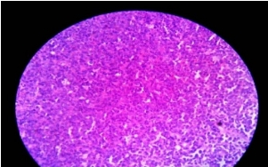
While imaging studies suggest the location of the lesion and help in narrowing the differential diagnosis, morphological assessment by fine needle aspiration cytology(FNAC) or histopathological examination is imperative prior to therapeutic intervention. Biopsy of mediastinal mass can be performed by variety of techniques ranging from FNAC to surgical procedures allowing resection or biopsy70. Although FNAC is a cost effective tool for the establishment of diagnosis of mediastinal masses, there is a complexity and technical difficulty associated with this technique owing to the narrow anatomic space of mediastinum. A major advantage of FNAC is that immediate cytological examination of specimen is possible71. Also, there are well known diagnostic pitfalls of this technique that limit its use2.
A thorough study of cytomorphology is extremely useful in reaching the correct diagnosis. The cytological smears may be composed of predominantly round cells, spindle cells or pleomorphic bizarre cells.
In certain situations no specific cell pattern may be noted. There are many other lesions in the mediastinum which are difficult to put in a specific group. These lesions consist of intrathoracic goitre, parathyroid adenomas, lipoma, bronchogenic cyst, paravertebral abscess etc72-74.
Sampling error is a major hurdle in the diagnosis of mediastinal lesions61. Tumours of the mediastinum may show necrosis, inflammation and fibrosis on FNAC smears. Needle may hit in the non-viable area and the quality of the material may be suboptimal. Tumour heterogeneity particularly in case of mixed germ cell tumours may also pose diagnostic challenge

Ancillary techniques

A battery of ancillary investigations may be needed for diagnosis of mediastinal tumours.
Immunocytochemistry:
It could be done either on alcohol fixed smears or cell block sections. Cell block is preferable for immunocytochemistry75-77.
Flow Cytometric Immunophenotyping:
This may be helpful in diagnosis and classification of non Hodgkin’s lymphomas77. It is advisable to have a panel of immunostain on suspected case of NHL.

Electron Microscopy:

Electron microscopy may be helpful in diagnosing the different mediastinal tumors78. It may be particularly helpful diagnosing metastatic squamous cell carcinoma, neuroblastoma or thymomas.

Biochemical Estimation:

Raised level of alpha feto protein may be helpful in endodermal sinus tumours. Similarly, high level of HCG is helpful in diagnosis of choriocarcinomas.

Microbial Culture and PCR:

A portion of the aspirate could be kept for microbial culture and PCR test like CBNAAT for tuberculosis in presumptive tuberculosis patient79.

Classification of mediastinal masses

Mediastinal masses are considered primary, that is, arising solely from structures within the mediastinum, or secondary, usually as metastatic disease from intrathoracic or extrathoracic malignancy. Mediastinal masses can be classified into malignant and benign which can be further divided into infectious and non infectious.
Numerous classification systems have been developed and used to varying degrees by anatomists, surgeons, and radiologists. The most commonly used scheme in clinical practice is the Shields classification system, whereas the traditional Fraser and Paré, Felson, Heitzman, Zylak, and Whitten models are used in radiologic practice8-14.
The most practical clinical classification of primary mediastinal masses groups together lesions that are characteristically found in the anterior, middle or posterior mediastinal compartments (table:1)24, with the recognition that such a simplified scheme overlooks the fact that mediastinal masses will not necessarily respect anatomic borders. Division of the mediastinum into specific compartments has traditionally been valuable in the identification, characterization, and management of various mediastinal abnormalities. Masses found within any of the mediastinal compartments may be due to lesions more commonly found in another mediastinal compartment or due to those that have extended from another area in the mediastinum.

Table: 1 Classification of Mediastinal Mass as per location

Anterior
Mediastinum
Middle
Mediastinum
Posterior
Mediastinum
Thymic lesions
Thymoma
Carcinoma
Hyperplasia
Cysts
Thymolipoma
Lymphadenopa
thy
Granulomatous
inflammation
Metastasis
Castleman’s
disease
Neurogenic
tumours
Germ cell
tumours
Teratoma
Seminoma
Non
seminomatous
germ cell
tumours
Embryonal cell
carcinoma
Choriocarcinoma
Lymphoma Meningocele
Lymphoma
Hodgkin
lymphoma
Non Hodgkin
lymphoma
Developmental
cyst
Pericardial cyst
Foregut
duplication cyst
Bronchogenic
cyst
Enteric cyst
Oesophageal
lesions
Carcinoma
Diverticula
Thyroid
neoplasm
Vascular
enlargements
Diaphragma
tic hernia
[bochdalek]
Parathyroid
neoplasm
Diaphragmatic
hernia[hiatus]
Miscellaneo
us
Mesenchymal
tumours
Lipoma
Fibroma
Lymphangioma
Haemangioma
Mesothelioma
Sarcoma
Diaphragmatic
hernia[morgagni]
Nut midline
carcinoma

Anterior Mediastinal Mass

In a patient with an anterior mediastinal mass, it is often possible to make a strong provisional diagnosis based upon clinical evaluation and imaging data. The common anterior mediastinal masses are thymomas, lymphoma and germ cell tumors80. In an individual younger than 40 years, lymphoma is the most likely diagnosis and the presence of palpable lymphadenopathy further increases the level of suspicion. In contrast to lymphoma, thymic neoplasm is very uncommon before the fourth decade of life. The presence of a paraneoplastic syndrome associated with an anterior mediastinal mass essentially clinches the diagnosis of thymoma. The majority of GCTs (benign or malignant) are diagnosed in the second or third decade of life. Whereas patients with a thymoma often have an indolent presentation, patients with a lymphoma or a malignant GCT often have a rapid onset of symptoms. Autoantibodies to the acetylcholine receptor (anti-AChR) should be measured as their presence is virtually diagnostic of myasthenia gravis, even if the patient is without obvious symptoms.
Although imaging is most often not diagnostic of the specific type of tumour, some imaging features can be pathognomonic. For example, the finding of a well-encapsulated lesion in the anterior mediastinum containing several tissue elements – calcium, fat, fluid – is essentially diagnostic of a mature teratoma.
Routine biopsy should not be endorsed, because of the potential risk of tumour spread. Well-encapsulated lesions believed not to be lymphoma are often resected for both diagnosis and treatment, without a preceding biopsy. Conversely, for locally invasive or frankly unresectable anterior mediastinal masses, a biopsy should often be performed as such lesions may represent lymphoma, aggressive thymomas that could benefit from neoadjuvant treatment. Once a clinical diagnosis of thymoma is made, the goal is to proceed directly to resection without preliminary biopsy, as these tumours have a predilection for local recurrence once the thymic capsule has been violated.
Surgical extirpation is the mainstay of treatment for mature GCTs, and biopsy is not indicated for these lesions if they have the characteristic imaging characteristics. Malignant GCTs, on the other hand, are treated primarily with chemotherapy, radiotherapy, or both. In situations where these lesions are suspected but tumour markers (AFP and β-HCG) are not markedly elevated, biopsy should be performed81.

Thymic Neoplasm

Thymic tumours are the most common primary tumours of the anterior mediastinum, constituting 30% to 50% of all the masses in this location; thymomas are responsible for most of these 82-84. Most patients are adults older than 40 years, with equal sex predilection 82, 83.
The Definitions of World Health Organization Classification of Thymic Epithelial Tumours[table: 2]24
Shamji F et al and Cohen DJ et al stated that approximately 50% of patients with thymoma are asymptomatic at the time of diagnosis85, 86. At diagnosis, symptoms due to tumour related syndromes or, more commonly, myasthenia gravis are present in up to one half of patients87, 88.
Conversely, Osserman KE et al stated that only 15% of patients with myasthenia gravis have a thymoma89. All patients suspected to have thymoma, even if they are asymptomatic, should have a serum antiacetylcholine (Ach) receptor antibody level examined to exclude myasthenia gravis90, 91.
Most thymomas arise in the upper anterior mediastinum but may project into the adjacent middle or posterior mediastinum25. They vary in size from very small to larger than 20 cm in diameter. Thymomas are usually well-defined, with spherical or lobulated borders92. Only the larger tumours are visible on plain radiograph25. On CT, encapsulated thymomas can appear as a homogeneous or heterogeneous soft tissue mass, depending on the presence of haemorrhage, necrosis, or cyst formation92. Other thymic neoplasm include thymic carcinoma, thymic carcinoid, and thymolipoma; the last two are very rare.
Table: 2 World Health Organization (WHO) Histological Classification System

A A tumour comprised of a homogenous population of neoplastic epithelial cells with spindle/oval shape, lacking nuclear atypia, and accompanied by few or no nonneoplastic lymphocytes
AB A tumour in which foci with the features of type A thymoma are admixed with foci rich in lymphocytes: the segregation of two patterns can be sharp or indistinct
B1 A tumour that resembles the normal functional thymus in that it combines large expanses with an appearance practically indistinguishable from that of normal thymic cortex with areas resembling thymic medulla
B2 A tumour in which the neoplastic epithelial component appears as scattered plump cells with vesicular nuclei and distinct nucleoli
among a heavy population of lymphocytes; perivascular spaces are common
B3 A tumour comprised predominantly of epithelial cells with a round or polygonal shape and exhibiting mild atypia admixed with a minor component of lymphocytes; foci of squamous metaplasia and perivascular spaces are common
C Thymic tumour exhibiting clear cut cytological atypia and a set of cytoarchitectural features no longer specific to the thymus ; but rather analogous to those seen in carcinomas of the other organs; type C thymomas lack immature lymphocytes ; whatever lymphocytes may be present are mature and usually admixed with plasma cells.

Lymphoma

Patients with Hodgkin lymphoma typically present with cervical or supraclavicular lymphadenopathy93. Patients with Hodgkin lymphoma who present with mediastinal involvement are usually younger than those who present without mediastinal disease (29 years versus 38 years) 96. Most patients with Hodgkin lymphoma who have an abnormal chest radiograph have bilateral asymmetric nodal disease97. The prevascular and paratracheal nodes are the most commonly affected nodes98 and only 15% of patients with intrathoracic Hodgkin lymphoma have enlargement of a single lymph node group94. Bulky nodal mediastinal disease can be caused by both Hodgkin lymphoma and Non Hodgkin lymphoma [NHL] 93, 99. NHL involves the anterior mediastinum less frequently than Hodgkin lymphoma95. Also, NHL has a greater tendency to non contiguously spread to the middle and posterior mediastinum94, 100.

Middle Mediastinal Masses

Mediastinal lymphadenopathy is the most common among middle mediastinal masses. The most common primary masses of the middle mediastinum are lymphoma and Developmental cysts. Cysts comprise 12% to 20% of all primary mediastinal masses and are found chiefly in the middle compartment of the mediastinum. These cysts comprise congenital foregut cysts, bronchogenic cysts, neurenteric cysts, and pericardial cysts81.

Mediastinal lymphadenopathy

Mediastinal lymph node enlargement is most often due to lymphoma102, metastatic cancer101, granulomatous inflammation such as that caused by sarcoidosis or infection. Infection should be considered when the adenopathy is associated with a pulmonary opacity. Tuberculosis is a notable cause of mediastinal adenopathy that can mimic sarcoidosis or malignancy and should be suspected in a host with known tuberculosis risk factors, such as recent known exposure or residence in an endemic area103.
Fungal infections that cause granulomas, particularly histoplasmosis, may present with mediastinal and hilar adenopathy in the absence of a pulmonary opacity104.Numerous less common causes of mediastinal adenopathy are described, including Castleman’s disease or angiofollicular lymphoid hyperplasia.

Tuberculosis

Tuberculous lymphadenitis is another important consideration for middle mediastinal mass. Tuberculosis (TB) is responsible for up to 43% of all of peripheral lymphadenopathy in the developing world. In the United States, 5.4% of all TB cases are extrapulmonary, and 31% of these are lymphatic131. Total estimated incidence of tuberculous lymphadenopathy was 30.8 per 100,000 population in India79. The cervical region is most frequently involved, but mediastinal involvement occurs in approximately 27% of cases. Tuberculous mediastinal lymphadenopathy presenting with dysphagia or oesophageal perforation has been reported129,130. Presumptive tuberculous mediastinal lymphadenopathy should be suspected in patients with cough, fever, shortness of breath, weight loss or night sweats who have hilar widening on chest X-ray and/or mediastinal lymphadenopathy on chest CT79.

Developmental cysts:

Developmental cysts of various sorts comprise 10% to 20% of all mediastinal masses105. Most can be identified as bronchogenic, enteric, or pericardial. Bronchogenic and enteric cysts are often referred to as foregut duplication cysts because of their origin from aberrant portions of the ventral and dorsal foregut, respectively. Bronchogenic cysts are found near large airways, often just posterior to the carina, although they may be attached to the oesophagus or even lie inside the pericardial space4. Developmental cysts can usually be identified by CT or ultrasonography, and the diagnosis may be confirmed by aspiration cytology. MRI is valuable for confirming the cystic nature of these lesions.

Posterior Mediastinal Mass

Neurogenic tumours are the most common neoplasm of the posterior mediastinum, collectively representing 12% to 21% of all mediastinal masses and occurring almost exclusively (95%) in the posterior compartment106. Neurogenic tumours are categorized into three groups based on the neurogenic tissue of origin. Neoplasms arising from the nerve sheath include schwannoma, neurofibroma, and malignant nerve sheath tumours. Nerve sheath tumours are the most common neurogenic tumours found in the adult. Neoplasms arising from the sympathetic ganglion include ganglioneuroma, ganglioneuroblastoma, and neuroblastoma and these tumours are more commonly found in children. Neoplasms arising in parasympathetic ganglia include paraganglioma and chemodectoma, both of which occur in the posterior mediastinum and are exceedingly rare. Approximately 98% of neurogenic mediastinal tumours in adults are benign, whereas it is estimated that more than 50% of neurogenic tumours in children are malignant24.

Classification of Neuroginc tumours:

Neoplasm arising from peripheral nerves
Neurofibroma
Neurilemoma (schwannoma)
Neurosarcoma
Neoplasm arising from sympathetic ganglia
Ganglioneuroma
Ganglioneuroblastoma
Neuroblastoma
Neoplasm arising from paraganglionic tissue
Pheochromocytoma
Paraganglioma (chemodectoma)

Peripheral Nerve Tumours

Schwannomas are the most common mediastinal neurogenic tumours, being responsible for 50% of mediastinal neurogenic tumours in adults107. Mediastinal benign schwannoma originates from Schwann cells; both schwannoma and solitary neurofibroma affect patients of both sexes predominantly in their third and fourth decade of life. Most patients are asymptomatic and 30% to 45% of neurofibromas occur in patients with neurofibromatosis107. On imaging studies, neurofibromas and schwannomas are generally well circumscribed homogeneous or heterogeneous, spherical, lobulated paraspinous masses108

Malignant tumour of nerve sheath origin

These are rare spindle cell sarcomas that typically arise from a simple or plexiform neurofibroma (well defined, non encapsulated tumour that usually infiltrates along an entire nerve trunk or plexus) and approximately 50% occur in patients with neurofibromatosis. These tumours are typically seen on CT as spherical well demarcated posterior mediastinal masses109.

Conclusion

The diagnosis of the mediastinal tumours requires a combination of clinical, radiological and pathological information. Nowadays availability of advanced imaging methods are more precise to determine the location of masses and also helps to choose the biopsy techniques with less invasive and more yield to know the exact nature of mass. Advent of newer techniques like EBUS and EUS has made tissue sampling from these sites easier5. To choose the Accurate and reliable diagnostic procedures, we should have wide knowledge about anatomical, clinical, radiological features, differential diagnosis of mediastinal mass based on location and about various biopsy techniques available.

Reference

1. Wychulis A. Surgical treatment of mediastinal tumors. A 40 year experience. J ThoracCardiovasc Surg. 1971;62:379-92.
2. Shabb NS, Fahl M, Shabb B, Haswani P, Zaatari G. Fine‐needle aspiration of the mediastinum: A clinical, radiologic, cytologic, and histologic study of 42 cases. Diagnostic cytopathology. 1998 Dec;19(6):428-36.
3. Wright CD, Mathisen DJ. Mediastinal tumors: diagnosis and treatment. World journal of surgery. 2001 Feb 1;25(2):204-9.
4. Silverman NA, Sabiston Jr DC. Mediastinal masses. Surgical Clinics of North America. 1980 Aug 1;60(4):757-77.
5. Roux BI, Kallichurum S, Shama DM. Mediastinal cysts and tumors. Current problems in surgery. 1984 Nov 1;21(11):6-77.
6. Cohen AJ, Thompson L, Edwards FH, Bellamy RF. Primary cysts and tumors of the mediastinum. The Annals of thoracic surgery. 1991 Mar 1;51(3):378-86.
7. Williams PL, Warwick R, Dyson M, Bannister LH. Splanchnology. In: Gray’s anatomy. 37th ed. New York, NY: Churchill Livingstone, 1989;1245–1475.
8. Shields TW. Primary tumours and cysts of the mediastinum. In: Shields TW, ed. General thoracic surgery. Philadelphia, Pa: Lea &Febiger, 1983; 927–954.
9. Fraser RS, Müller NL, Colman N, Paré PD, eds. The mediastinum.In: Fraser and Paré’s diagnosis of diseases of the chest. 4th ed. Philadelphia, Pa: Saunders, 1999; 196–234. Book ref
10. Fraser RG, Paré PA, eds. The normal chest. In: Diagnosis of diseases of the chest. 2nd ed. Philadelphia, Pa: Saunders, 1977; 1–183.
11. Felson B. Chest roentgenology. Philadelphia, Pa: Saunders, 1973.
12. Heitzman ER. The mediastinum. 2nd ed. New York, NY: Springer-Verlag, 1988.
13. Zylak CJ, Pallie W, Jackson R. Correlative anatomy and computed tomography: a module on the mediastinum. Radiographics. 1982 Nov;2(4):555-92.
14. Whitten CR, Khan S, Munneke GJ, Grubnic S. A diagnostic approach to mediastinal abnormalities. Radiographics. 2007 May;27(3):657-71.
15. Sharma P, Jha V, Kumar N, Kumar R, Mandal A. Clinicopathological analysis of mediastinal masses: a mixed bag of non-neoplastic and neoplastic etiologies. Turkish Journal of Pathology. 2017 Jan 1;33(1):037-46.
16. Dixit R, Shah NS, Goyal M, Patil CB, Panjabi M, Gupta RC, Gupta N, Harish SV. Diagnostic evaluation of mediastinal lesions: Analysis of 144 cases. Lung India: official organ of Indian Chest Society. 2017 Jul;34(4):341
17. Williams PL, Warwick R, Dyson M, Bannister LH. Splanchnology. In: Gray’s anatomy. 37th ed. New York, NY: Churchill Livingstone, 1989; 1245–1475.
18. Naruke T, Suemasu K, Ishikawa S. Lymph node mapping and curability at various levels of metastasis in resected lung cancer. The Journal of thoracic and cardiovascular surgery. 1978 Dec;76(6):832-9.
19. Donahue JM, Nichols FC. Primary mediastinal tumors and cysts and diagnostic investigation of mediastinal masses. Shields TW, Lo Cicero III J, Reed CE, Feins RH, editors. General Thoracic Surgery. Philadelphia: Lippincott Williams & Wilkins. 2009:2489-94.
20. Davis RD, Oldham HN, Sabiston DC. Primary cysts and neoplasms of the mediastinum: recent changes in clinical presentation, methods of diagnosis, management, and results. The Annals of thoracic surgery. 1987 Sep 1;44(3):229-37.
21. Henschke CI, Lee IJ, Wu N, Farooqi A, Khan A, Yankelevitz D, Altorki NK. CT screening for lung cancer: prevalence and incidence of mediastinal masses. Radiology. 2006 May;239(2):586-90.
22. Aroor AR, Rama Prakasha S, Seshadri S, Teerthanath S, Raghuraj U. A study of clinical characteristicsof mediastinal mass. Journal of clinical and diagnostic research: JCDR. 2014 Feb;8(2):77.
23. Wilson LD, Detterbeck FC, Yahalom J. Superior vena cava syndrome with malignant causes. New England Journal of Medicine. 2007 May 3;356(18):1862-9.
24. Murray JF, Nadel JA, editors. Textbook of respiratory medicine. 6th ed. Philadelphia: W.B. Saunders; 2016: 1478–96. Book ref
25. Armstrong P. Mediastinal and hilar disorders. In: Armstrong P, Wilson AG, Dee P, Hansell DM, editors. Imaging of diseases of the chest. 3rd ed. London: Mosby; 2000: 789–892. Book ref
26. Ahn JM, Lee KS, Goo JM, Song KS, Kim SJ, Im JG. Predicting the histology of anterior mediastinal masses: comparison of chest radiography and CT. Journal of thoracic imaging. 1996;11(4):265-71.
27. Naidich DP, Zerhouni EA, Siegelman SS, Kuhn JP. Computed tomography and magnetic resonance of the thorax.
28. Silvestri GA, Gonzalez AV, Jantz MA, Margolis ML, Gould MK, Tanoue LT, Harris LJ, Detterbeck FC. Methods for staging non-small cell lung cancer: diagnosis and management of lung cancer: American College of Chest Physicians evidence-based clinical practice guidelines. Chest. 2013 May 1;143(5):e211S-50S.
29. Gould MK, Kuschner WG, Rydzak CE, Maclean CC, Demas AN, Shigemitsu H, Chan JK, Owens DK. Test performance of positron emission tomography and computed tomography for mediastinal staging in patients with non–small-cell lung cancer: a metaanalysis. Annals of internal medicine. 2003 Dec 2;139(11):879-92.
30. Ratto GB, Frola C, Cantoni S, Motta G. Improving clinical efficacy of computed tomographic scan in the preoperative assessment of patients with non-small cell lung cancer. The Journal of thoracic and cardiovascular surgery. 1990 Mar;99(3):416-25.
31. McLoud TC, Bourgouin PM, Greenberg RW, Kosiuk JP, Templeton PA, Shepard JA, Moore EH, Wain JC, Mathisen DJ, Grillo HC. Bronchogenic carcinoma: analysis of staging in the mediastinum with CT by correlative lymph node mapping and sampling. Radiology. 1992 Feb;182(2):319-23.
32. Tomiyama N, Honda O, Tsubamoto M, Inoue A, Sumikawa H, Kuriyama K, Kusumoto M, Johkoh T, Nakamura H. Anterior mediastinal tumors: diagnostic accuracy of CT and MRI. European journal of radiology. 2009 Feb 1;69(2):280- 8.
33. Ackman JB, Wu CC. MRI of the thymus. American Journal of Roentgenology. 2011 Jul;197(1):W15-20.
34. Ackman JB. MR imaging of mediastinal masses. Magnetic Resonance Imaging Clinics. 2015 May 1;23(2):141-64.
35. Carter BW, Betancourt SL, Benveniste MF. MR imaging of mediastinal masses. Topics in Magnetic Resonance Imaging. 2017 Aug 1;26(4):153-65.
36. Inaoka T, Takahashi K, Mineta M, Yamada T, Shuke N, Okizaki A, Nagasawa K, Sugimori H, Aburano T. Thymic hyperplasia and thymus gland tumors: differentiation with chemical shift MR imaging. Radiology. 2007 Jun;243(3):869-76.
37. Takahashi K, Al‐Janabi NJ. Computed tomography and magnetic resonance imaging of mediastinal tumors. Journal of magnetic resonance imaging. 2010 Dec;32(6):1325-39.
38. Kubota K, Yamada S, Kondo T, Yamada K, Fukuda H, Fujiwara T, Ito M, Ido T. PET imaging of primary mediastinal tumours. British journal of cancer. 1996 Apr;73(7):882.
39. Tatci E, Ozmen O, Dadali Y, Biner IU, Gokcek A, Demirag F, Incekara F, Arslan N. The role of FDG PET/CT in evaluation of mediastinal masses and neurogenic tumors of chest wall. International journal of clinical and experimental medicine. 2015;8(7):11146.
40. Lee BE, von Haag D, Lown T, Lau D, Calhoun R, Follette D. Advances in positron emission tomography technology have increased the need for surgical staging in non–small cell lung cancer. The Journal of thoracic and cardiovascular surgery. 2007 Mar 1;133(3):746-52.
41. National Comprehensive Cancer Network: NCCN clinical practice guidelines in oncology: lung cancer screening. Version 1.2013 <http://www.nccn.org/professionals/physician_gls/pdf/lung_screening.pdf>.Accessed June 12, 2013.
42. Puri V, Meyers BF. Utility of positron emission tomography in the mediastinum: moving beyond lung and esophageal cancer staging. Thoracic Surgery Clinics. 2009 Feb 1;19(1):7-15.
43. Sung YM, Lee KS, Kim BT, Choi JY, Shim YM, Chin AY. 18F-FDG PET/CT of thymic epithelial tumors: usefulness for distinguishing and staging tumor subgroups. Journal of Nuclear Medicine. 2006 Oct 1;47(10):1628-34.
44. Treglia G, Sadeghi R, Giovanella L, Cafarotti S, Filosso P, Lococo F. Is 18F-FDG PET useful in predicting the WHO grade of malignancy in thymic epithelial tumors? A meta-analysis. Lung Cancer. 2014 Oct 1;86(1):5-13.
45. Carter BW, Benveniste MF, Madan R, Godoy MC, de Groot PM, Truong MT, Rosado-de-Christenson ML, Marom EM. ITMIG classification of mediastinal compartments and multidisciplinary approach to mediastinal masses. Radiographics. 2017 Jan 27;37(2):413-36.
46. Shure D, Fedullo PF. The role of transcarinal needle aspiration in the staging of bronchogenic carcinoma. Chest. 1984 Nov 1;86(5):693-6.
47. Schenk DA, Bower JH, Bryan CL, Currie RB, Spence TH, Duncan CA, Myers DL, Sullivan WT. Transbronchial needle aspiration staging of bronchogenic carcinoma. American Review of Respiratory Disease. 1986 Jul;134(1):146-8.
48. Rintoul RC, Tournoy KG, El Daly H, Carroll NR, Buttery RC, Van Kralingen K, van Meerbeeck JP, Rabe KF, Annema JT. EBUS-TBNA for the clarification of PET positive intra-thoracic lymph nodes—an international multi-centre experience. Journal of Thoracic Oncology. 2009 Jan 1;4(1):44-8.
49. Herth FJ, Becker HD, Ernst A. Ultrasound-Guided Transbronchial Needle Aspiration*: An Experience in 242 Patients. Chest. 2003 Feb 1;123(2):604-7
50. Yasufuku K, Nakajima T, Motoori K, Sekine Y, Shibuya K, Hiroshima K, Fujisawa T. Comparison of endobronchial ultrasound, positron emission tomography, and CT for lymph node staging of lung cancer. Chest. 2006 Sep 1;130(3):710-8.
51. Yasufuku K, Nakajima T, Fujiwara T, Chiyo M, Iyoda A, Yoshida S, Suzuki M, Sekine Y, Shibuya K, Yoshino I. Role of endobronchial ultrasound-guided transbronchial needle aspiration in the management of lung cancer. General thoracic and cardiovascular surgery. 2008 Jun 1;56(6):268-76.
52. Tremblay A, Stather DR, MacEachern P, Khalil M, Field SK. A randomized controlled trial of standard vs endobronchial ultrasonography-guided transbronchial needle aspiration in patients with suspected sarcoidosis. Chest. 2009 Aug 1;136(2):340-6.
53. Navani N, Lawrence DR, Kolvekar S, Hayward M, McAsey D, Kocjan G, Falzon M, Capitanio A, Shaw P, Morris S, Omar RZ. Endobronchial ultrasound–guided transbronchial needle aspiration prevents mediastinoscopies in the diagnosis of isolated mediastinal lymphadenopathy: a prospective trial. American journal of respiratory and critical care medicine. 2012 Aug 1;186(3):255-60.
54. Krasnik M. Endoscopic transesophageal and endoscopic transbronchial real-time ultrasound-guided biopsy. Respiration. 2003;70(3):293-8.
55. Gress FG, Savides TJ, Sandler A, Kesler K, Conces D, Cummings O, Mathur P, Ikenberry S, Bilderback S, Hawes R. Endoscopic ultrasonography, fine-needle aspiration biopsy guided by endoscopic ultrasonography, and computed tomography in the preoperative staging of non-small-cell lung cancer: a comparison study. Annals of internal medicine. 1997 Oct 15;127(8_Part_1):604-12.
56. Akamatsu H, Terashima M, Koike T, Takizawa T, Kurita Y. Staging of primary lung cancer by computed tomography-guided percutaneous needle cytology of mediastinal lymph nodes. The Annals of thoracic surgery. 1996 Aug 1;62(2):352-5.
57. Moinuddin SM, Lee LH, Montgomery JH. Mediastinal needle biopsy. American journal of roentgenology. 1984 Sep 1;143(3):531-2.
58. Protopapas Z, Westcott JL. Transthoracic hilar and mediastinal biopsy. Radiologic Clinics of North America. 2000 Mar 1;38(2):281-91.
59. Wiersema MJ, Vazquez-Sequeiros E, Wiersema LM. Evaluation of mediastinal lymphadenopathy with endoscopic US-guided fine-needle aspiration biopsy. Radiology. 2001 Apr;219(1):252-7.
60. Gupta S, Seaberg K, Wallace MJ, Madoff DC, Morello Jr FA, Ahrar K, Murthy R, Hicks ME. Imaging-guided percutaneous biopsy of mediastinal lesions: different approaches and anatomic considerations. Radiographics. 2005 May;25(3):763-86.
61. Singh HK, Silverman JF, Powers CN, Geisinger KR, Frable WJ. Diagnostic pitfalls in fine‐needle aspiration biopsy of the mediastinum. Diagnostic cytopathology. 1997 Aug;17(2):121-6.
62. Assaad MW, Pantanowitz L, Otis CN. Diagnostic accuracy of image‐guided percutaneous fine needle aspiration biopsy of the mediastinum. Diagnostic cytopathology. 2007 Nov;35(11):705-9.
63. Kelemen III JJ, Naunheim KS. Minimally invasive approaches to mediastinal neoplasms. InSeminars in Thoracic and Cardiovascular surgery 2000 Oct 1 (Vol. 12, No. 4, pp. 301-306). WB Saunders.
64. Ginsberg RJ. Evaluation of the mediastinum by invasive techniques. The Surgical clinics of North America. 1987 Oct;67(5):1025-35.
65. Vallières E, Pagé A, Verdant A. Ambulatory mediastinoscopy and anterior mediastinotomy. The Annals of thoracic surgery. 1991 Nov 1;52(5):1122-6.
66. Prinz RA, Lonchyna V, Carnaille B, Wurtz A, Proye C. Thoracoscopic excision of enlarged mediastinal parathyroid glands. Surgery. 1994 Dec 1;116(6):999-1005
67. Hazelrigg SR, Landreneau RJ, Mack MJ, Acuff TE. Thoracoscopic resection of mediastinal cysts. The Annals of thoracic surgery. 1993 Sep 1;56(3):659-60.
68. Sugarbaker DJ. Thoracoscopy in the management of anterior mediastinal masses. The Annals of thoracic surgery. 1993 Sep 1;56(3):653-6.
69. Demmy TL, Krasna MJ, Detterbeck FC, Kline GG, Kohman LJ, DeCamp Jr MM, Wain JC. Multicenter VATS experience with mediastinal tumors. The Annals of thoracic surgery. 1998 Jul 1;66(1):187-92
70. Morrissey B, Adams H, Gibbs AR, Crane MD. Percutaneous needle biopsy of the mediastinum: review of 94 procedures. Thorax. 1993 Jun 1;48(6):632-7.
71. Rendina EA, Venuta F, De Giacomo T, Ciccone AM, Moretti MS, Ibrahim M, Coloni GF. Biopsy of anterior mediastinal masses under local anesthesia. The Annals of thoracic surgery. 2002 Nov 1;74(5):1720-3.
72. Strollo DC, de Christenson Lt ML, Jett JR. Primary mediastinal tumors. Part 1*: tumors of the anterior mediastinum. Chest. 1997 Aug 1;112(2):511-22.
73. Clark OH. Mediastinal parathyroid tumors. Archives of Surgery. 1988 Sep 1;123(9):1096-100.
74. Wong NA, Mihai R, Sheffield EA, Calder CJ, Farndon JR. Imprint cytology of parathyroid tissue in relation to other tissues of the neck and mediastinum. Actacytologica. 2000;44(2):109-13.
75. Beckstead JH. Alkaline phosphatase histochemistry in human germ cell neoplasms. The American journal of surgical pathology. 1983 Jun;7(4):341-9.
76. Yatabe Y, Mitsudomi T, Takahashi T. TTF-1 expression in pulmonary adenocarcinomas. The American journal of surgical pathology. 2002 Jun 1;26(6):767-73.
77. Dey P, Amir T, Al Jassar A, Al Shemmari S, Jogai S. Combined applications of fine needle aspiration cytology and Flow cytometricimmunphenotyping for diagnosis and classification of non Hodgkin Lymphoma. Cytojournal. 2006;3:24.
78. Neill JS, Silverman JF. Electron microscopy of fine‐needle aspiration biopsies of the mediastinum. Diagnostic cytopathology. 1992 May;8(3):272-7.
79. Index TB. Guidelines. Guidelines on extra-pulmonary tuberculosis for India. World Health Organization. 2016.
80. Hoerbelt R, Keunecke L, Grimm H, Schwemmle K, Padberg W. The value of a noninvasive diagnostic approach to mediastinal masses. The Annals of thoracic surgery. 2003 Apr 1;75(4):1086-90.
81. Bryan M. Burt,Joseph B. ShragerBenign and Malignant Neoplasm of the Mediastinum.Fishman’s Pulmonary Diseases and Disorders, fifth edition.82. Book ref
82. Lewis JE, Wick MR, Scheithauer BW, Bernatz PE, Taylor WF. Thymoma. A clinicopathologic review. Cancer. 1987 Dec 1;60(11):2727-43.
83. MORGENTHALER TI, BROWN LR, COLBY TV, HARPER Jr CM, COLES DT. Thymoma. InMayo Clinic Proceedings 1993 Nov 1 (Vol. 68, No. 11, pp. 1110-1123). Elsevier.
84. Mullen B, Richardson JD. Primary anterior mediastinal tumors in children and adults. The Annals of thoracic surgery. 1986 Sep 1;42(3):338-45.
85. Shamji F, Pearson FG, Todd TR, Ginsberg RJ, Ilves R, Cooper JD. Results of surgical treatment for thymoma. The Journal of thoracic and cardiovascular surgery. 1984 Jan;87(1):43-7.
86. Cohen DJ, Ronnigen LD, Graeber GM, Deshong JL, Jaffin J, Burge JR, Zajtchuk R. Management of patients with malignant thymoma. The Journal of thoracic and cardiovascular surgery. 1984 Feb;87(2):301-7.
87. Silverman NA, Sabiston Jr DC. Mediastinal masses. Surgical Clinics of North America. 1980 Aug 1;60(4):757-77.
88. Gerein AN, Srivastava SP, Burgess J. Thymoma: a ten year review. The American Journal of Surgery. 1978 Jul 1;136(1):49-53.
89. Osserman KE. Studies in myasthenia gravis-review of twenty year experience in over 1200 patients. Mt Sinai J Med. 1971;38:497-537.
90. Elias SB, Appel SH, Lennon VA, Jones G, Howard F, Elveback L, Drachman DB, Adams RN, Josifek LF, Self SG. Autoantibodies to acetylcholine receptors in myasthenia gravis. New England Journal of Medicine. 1983 Feb 17;308(7):402-3.
91. HOWARD JR FM, Lennon VA, Finley JO, Matsumoto J, Elveback LR. Clinical Correlations of Antibodies That Bind, Block, or Modulate Human Acetylcholine Receptors in Myasthenia Gravis a. Annals of the New York Academy of Sciences. 1987 Aug;505(1):526-38.
92. Rosado-de-Christenson ML, Galobardes J, Moran CA. Thymoma: radiologic-pathologic correlation. Radiographics. 1992 Jan;12(1):151-68.
93. Strickler JG, Kurtin PJ. Mediastinal lymphoma. In Seminars in diagnostic pathology 1991 Feb 1 (Vol. 8, No. 1, pp. 2-13). Elsevier.
94. Filly R, Blank N, Castellino RA. Radiographic distribution of intrathoracic disease in previously untreated patients with Hodgkin’s disease and nonHodgkin’s lymphoma. Radiology. 1976 Aug;120(2):277-81.
95. Costello P, Jochelson M. Lymphoma of the mediastinum and lung. Radiology: diagnosis, imaging, intervention. 1996;1:1-3.
96. Schomberg PJ, Evans RG, O’connell MJ, White WL, Banks PM, Ilstrup DM, Earle JD. Prognostic significance of mediastinal mass in adult Hodgkin’s disease. Cancer. 1984 Jan 15;53(2):324-8.
97. Mann RB, Jaffe ES, Berard CW. Malignant lymphomas– a conceptual understanding of morphologic diversity. A review. The American journal of pathology. 1979 Jan;94(1):105.
98. Castellino RA, Blank N, Hoppe RT, Cho C. Hodgkin disease: contributions of chest CT in the initial staging evaluation. Radiology. 1986 Sep;160(3):603-5.
99. Hopper KD, Diehl LF, Lesar M, Barnes M, Granger E, Baumann J. Hodgkin disease: clinical utility of CT in initial staging and treatment. Radiology. 1988 Oct;169(1):17-22.
100. Ellert J, Kreel L. The role of computed tomography in the initial staging and subsequent management of the lymphomas. Journal of computer assisted tomography. 1980 Jun;4(3):368-91.
101. McLoud TC, Meyer JE. Mediastinal metastases. Radiologic clinics of North America. 1982 Sep;20(3):453.
102. Filly R, Blank N, Castellino RA. Radiographic distribution of intrathoracic disease in previously untreated patients with Hodgkin’s disease and nonHodgkin’s lymphoma. Radiology. 1976 Aug;120(2):277-81.
103. Medford AR, Agrawal S. Single bronchoscope combined endoscopic-endobronchial ultrasoundguided fine-needle aspiration for tuberculous mediastinal nodes. Chest. 2010 Nov 1;138(5):1274.
104. Wheat LJ, Conces D, Allen SD, Blue-Hnidy D, Loyd J. Pulmonary histoplasmosis syndromes: recognition, diagnosis, and management. InSeminars in respiratory and critical care medicine 2004 Apr (Vol. 25, No. 02, pp.129-144). Copyright© 2004 by Thieme Medical Publishers, Inc., 333 Seventh Avenue, New York, NY 10001, USA.
105. Cioffi U, Bonavina L, De Simone M, Santambrogio L, Pavoni G, Testori A, Peracchia A. Presentation and surgical management of bronchogenic and esophageal duplication cysts in adults. Chest. 1998 Jun 1;113(6):1492-6.
106. Moran CA, Walsh G, Suster S, Kaiser L. Thymomas II: a clinicopathologic correlation of 250 cases with a proposed staging system with emphasis on pathologic assessment. American journal of clinical pathology. 2012 Mar 1;137(3):451-61.
107. Reed JC, Hallet KK, Feigin DS. Neural tumors of the thorax: subject review from the AFIP. Radiology. 1978 Jan;126(1):9-17.
108. Aughenbaugh GL. Thoracic manifestations of neurocutaneous diseases. Radiologic clinics of NorthAmerica. 1984 Sep;22(3):741-56.
109. Coleman BG, Arger PH, Dalinka MK, Obringer AC, Raney BR, Meadows AT. CT of sarcomatous degeneration in neurofibromatosis. American Journal of Roentgenology. 1983 Feb 1;140(2):383-7.


735 views

“Localised Bronchiectasis” – Time to Look Inside?

By | volume3-issue1 | No Comments

699 views

Mobeen Quadri et al.: “Localised Bronchiectasis” – Time to look Inside?

Case Report

“Localised Bronchiectasis” – Time to Look Inside?

Mobeen Quadri1, P. Yugandhar2

1Postgraduate – Department of Respiratory Medicine, Asram Medical College, Eluru, Andhra Pradesh, India
2Prof & HOD – Department of Respiratory Medicine, Asram Medical College, Eluru, Andhra Pradesh, India

Abstract

Bronchiectasis is a well-known entity where the airways abnormally dilate, losing their natural function.The most common causes of non-cystic fibrosis bronchiectasis in the middle age group includesecondary immunodeficiency, aspiration, and allergic bronchopulmonary aspergillosis (ABPA). An obstructive foreign body is an uncommon cause of Bronchiectasis and is often a missed diagnosis in localized disease. Foreign bodies can be missed making the diagnosis and treatment more challenging, and hence foreign body bronchiectasis should be considered in-patients presenting with focal disease. Here we describe a patient with a retained foreign body for a long duration of more than three decades. It was discovered through fiberoptic bronchoscopy with no significant aspiration history and non- diagnostic imaging of the chest.
Keywords: Foreign body, Aspiration, Fiberoptic bronchoscopy, Bronchiectasis

Corresponding Author: Dr.P.Yugandhar , Prof & HOD – Department of Respiratory Medicine, Asram Medical College, Eluru,
Andhra Pradesh, India. Email:yugs@rediffmail.com

Introduction

Bronchiectasis is a clinical entity where the airways are permanently and abnormally dilated due to an insult. Bronchiectatic airways lose their function, including mucociliary clearance, which results in bacterial overgrowth and repeated infections. Many insults have been identified to cause damage to the airways. The most common causes in middle-aged patients are secondary immunodeficiency syndromes, allergic bronchopulmonary aspergillosis (ABPA), chronic aspiration, metastatic malignancy, and retained foreign body. Frequently the cause of Bronchiectasis remains

unknown. Here we present a case of focal Bronchiectasis caused by a long-standing foreign body.

Case Report

  • A 38-year-old female patient came to our OPD for the first time with cough and expectoration and SOB for two months.
  • She gave a history of similar complaints in the past and the patient denied a history of any aspiration when asked specifically.
  • O/E Vitals were normal and respiratory system shows left side NVBS, and right side with coarse inspiratory crackles present in infra-axillary and infrascapular areas.

How to cite this article: Mobeen Quadri1, P. Yugandhar, “Localised Bronchiectasis” – Time to Look Inside?, JAPT 2020; 3(1):34-37

  • Investigations like blood parameters were normal, sputum for AFB & CBNAAT was negative.
  • CXR PA view shows right mid & lower zone non-homogenous opacity, HRCT chest shows cystic bronchiectasis in right middle lobe & basal segments of right lower lobe with dependent air-fluid levels.
  • Diagnostic Fiberoptic Bronchoscopy was done because of unilateral lesions & persistent/recurrent symptoms to rule out endobronchial pathology. To our surprise,
    there was a 2 cm wide foreign body lodged longitudinally in the basal segmental bronchi of lowerlobe, partly occluding the lumen.
  • On probing with biopsy forceps, it was moving. Hence we decided to remove it by holding with cupped biopsy forceps in toto along with Bronchoscope. On cleaning the debris, it turned out to be a piece of Tea Sachet.
  • After the removal, she improved symptomatically, and a new interview allowed the patient to recall that if she had any aspiration episode in the past, She gave a history of choking episode nearly 30 years back after opening a tea sachet with her mouth to make tea. Parents revealed history of recurrent symptoms after that episode.
  • Immediately after removal, CXR has been done.
  • Follow up CXR &CT scan after seven months shows No Improvement.

Discussion

  • We have presented a case of non-cystic fibrosis bronchiectasis in a middle-aged woman due to retained foreign body, which was not apparent with history or imaging. This cause of bronchiectasis in adults has been reported in the past but is rare.
  • Pathophysiology of the disease includes infection and impaired drainageor impaired host defense system like in our patient. The ensuing host response system of mostly neutrophils, cytokines, reactive oxygen species, and proteases causes chronic inflammation of the airways with subsequent ulceration and airway dilation leading to bronchiectasis.
  • Foreign body aspiration in the Tracheo- bronchial tree commonly occurs in children however, in adults, it is uncommon and challenging to make a diagnosis because of non-specific symptoms and CXR findings.
  • In our case, the foreign body persisted for a long time (30yrs) with recurrent infections.
  • Diagnostic bronchoscopy should be done in all suspicious cases. In our case, bronchoscopy turned out to be therapeutic too, by removing the foreign body which is present at lowerlobe basal segmental bronchi.
  • The most common foreign bodies are bone fragments or Food matter & ideal locations are right intermediate and right basal bronchi.
  • Early intervention can avoid permanent sequelae, and complete resolution is seen, whereas delayed intervention will lead to permanent damage and require surgical intervention for symptomatic relief.

Reference

1. Mise K, Jurcev Savicevic A, Pavlov N, Jankovic S. Removal of tracheobronchial foreign bodies in adults using flexible bronchoscopy: experience 1995-2006. Surg Endosc 2009;23(6):1360-1364.
2. Blanco Ramos M, Botana-Rial M, García- Fontán E, Fernández-Villar A, Gallas Torreira M. Update in the extraction of airway foreign bodies in adults. J Thorac Dis 2016;8(11):3452-3456. doi: 10.21037/jtd.2016.11.32
3. Dikensoy O, Usalan C, Filiz A. Foreign body aspiration: clinical utility of flexible bronchoscopy. Postgrad Med J 2002;78:399-403.
4. Al-Majed SA, Ashour M, Al-Mobeireek AF, Al-Hajjaj MS, Alzeer AH,Al-Kattan K. Overlooked inhaled foreign bodies: late sequelae and the likelihood of recovery. Respir Med 1997;91(5):293 296.
5. Lin L, Lv L, Wang Y, et al. The clinical features of foreign body aspiration into the lower airway in geriatric patients. Clin Interv Aging 2014;9:1613-8.
6. Debeljak A, Sorli J, Music E, et al. Bronchoscopic removal of foreign bodies in adults: experience with 62 patients from 1974-1998. Eur Respir J 1999;14:792-5.
7. Palasamudram Shekar S, Bajarano P, Hadeh A, et al. (July 12, 2018) Case of Missing Plastic: Foreign Body Bronchiectasis. Cureus 10(7): e2974. DOI 10.7759/cureus.2974
8.Hsu AA. Endoscopic intervention of lower airway foreign matter in adults-a different perspective. J Thorac Dis 2015;7:1870-7.
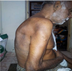
9. Sehgal IS, Dhooria S, Ram B, et al. Foreign Body Inhalation in the Adult Population: Experience of 25,998 Bronchoscopies and Systematic Review of the Literature. Respir Care 2015;60:1438-48.
10. Du Rand IA, Blaikley J, Booton R, et al. British Thoracic Society guideline for diagnostic flexible bronchoscopy in adults: accredited by NICE. Thorax 2013;68 Suppl
1:i1-i44.
11.Donado Un ̃a JR, de Miguel Poch E, Casado Lo ́pez ME, Alfaro AbreuJJ. Fiber optic bronchoscopy in extraction of tracheo-bronchial foreignbodies in adults. Arch
Bronconeumol 1998;34(2):76-81.
12. Soysal O, Kuzucu A, Ulutas H. Tracheobronchial foreign body aspiration:a continuing challenge. Otolaryngol Head Neck Surg 2006;135(2):223-226

379 views

A Rare Case Report of Anterior Mediastinal Paraganglioma Presenting as Superior Venacaval Obstruction

By | volume3-issue1 | No Comments

699 views

Keerthi Baskaran et al.: A Rare Case Report of Anterior Mediastinal Paraganglioma Presenting as Superior Venacaval Obstruction

Case Report

A Rare Case Report of Anterior Mediastinal Paraganglioma Presenting as Superior Venacaval Obstruction

Keerthi Baskaran1, Chitrakumar Ananthakrishnan2, Anandeswari Palanivelu3, Murugan Natrajan4

1Dr. Keerthi Baskaran MBBS., MD (Tuberculosis & Respiratory Medicine)
2Dr. Chitrakumar Ananthakrishnan MD (Tuberculosis & Respiratory Medicine)
3Dr. Anandeswari Palanivelu MD (Tuberculosis and Respiratory Medicine)
4Dr. Murugan Natrajan. MD (Tuberculosis and Respiratory Medicine)
Institute of Thoracic Medicine, Rajiv Gandhi Government General Hospital, Madras Medical College

Abstract

A 44 year old male, a chronic smoker presented with of hoarseness of voice for 5 years, loss of weight for 3 months, breathlessness for 10 days. Physical examination showed dilated veins over the anterior chest wall. CT chest revealed a mass extending from the lower part of the neck to the anterior mediastinum displacing and compressing the trachea and vascular structures. CT guided biopsy of the mass lesion led to the diagnosis of anterior mediastinal paraganglioma. Since the tumour was unresectable at the time of presentation, surgery was deferred and he underwent radiotherapy. In due course of time patient succumbed to the disease and expired 4 months after diagnosis. This case is being reported due to rarity of the disease. Mediastinal paraganglioma accounts for only about 2% of the paraganglioma, and we report a case of anterior mediastinal paraganglioma with superior venacaval obstruction which is exceptionally rare.
Keywords: Anterior mediastinal paraganglioma, Neuron specific enolase, CT guided biopsy, Superior vena caval obstruction syndrome
Key Messages: Anterior mediastinal paragangliomas are rare mediastinal tumours and should be kept in mind while evaluating any mediastinal mass. Complete surgical resection is the treatment of choice. This case is reported due to the rarity of occurrence of paraganglioma in the anterior mediastinum.

Corresponding Author: Dr. Keerthi Baskaran, Institute of Thoracic Medicine, Rajiv Gandhi Government General Hospital, Madras Medical College. Email:drkbs07@gmail.com

Introduction

Paragangliomas are unusual neoplasms that arise in various locations in our body. Mediastinal paraganglioma originate from para-aortic and paravertebral sympathetic chain ganglia and aortico-pulmonary paraganglia. Anterior mediastinal paragangliomas are extremely rare. Mostly the patients are asymptomatic and the diagnosis is incidental. Some cases present with hypertension, features

How to cite this article: Keerthi Baskaran, Chitrakumar Ananthakrishnan, Anandeswari Palanivelu, Murugan Natrajan, A Rare Case Report of Anterior Mediastinal Paraganglioma Presenting as Superior Venacaval Obstruction, JAPT 2020; 3(1):38-41

of catecholamine hypersecretion or features of mass effect on mediastinal structures. Complete resection of tumour is the treatment of choice.
Other modalities of treatment available for paragangliomas which are unresectable or having high risk of surgical complications are chemotherapy and radiotherapy. Here we are reporting a rare case of anterior mediastinal paraganglioma with superior venacaval obstruction.

Case History

A 44 year old male patient, a chronic smoker and an alcoholic with no pre-existing co- morbidities presented with complaints of hoarseness of voice for 5 years, which worsened over the past 3 months. He had loss of weight of about 6 kgs in the past 3 months, cough with sputum production and breathlessness for 10 days. He did not give any history of chest pain, hemoptysis, wheeze. He was a pest controller by occupation. On examination he had dilated veins over the left anterior chest wall. Patient was tachypenic at rest. Respiratory system examination revealed impaired to dull note on percussion of the sternal body, bilateral mammary areas. Diminished breath soundswere heard in bilateral mammary areas. Other major systems examination was normal. Chest roentgenogram was taken. (Fig 1 X-ray chest PA view, Left lateral view showing obliteration of retrosternal space). Routine blood investigations were within normal limits. ESR was 30 mm in 1⁄2 hour and 65 mm in 1 hour. Mantoux test and sputum AFB were negative. Sputum cytology for malignant/ atypical cells was negative. ECHO revealed the presence of moderate pericardial effusion. USG abdomen showed a rim of left pleural effusion. We proceeded with a CT chest (Fig 2 Hypodense lesion in the anterior mediastinum) which showed a well-defined hypodense lesion in the anterior mediastinum extending from the lower neck to anterior pericardium abutting the anterior chest wall, compressing and displacing the trachea and vascular structures. CT guided biopsy showed fragments of tumour tissue containing nests of small round to oval cells separated by richly vascular thin stroma. Cells and nuclei are uniform in size and shape separated focally by desmoplastic stroma with foci of calcification. Immunohistochemistry was found to be strongly positive for neuron specific enolase, weak to moderate activity for synaptophysin and chromogranin, no significant activity for Ki67 suggesting that it was paraganglioma of benign nature. Due to the close proximity to great vessels and high probability of complications on table, surgical resection of tumour was deferred and patient was advised to undergo radiation therapy. Patient succumbed to the disease 4 months after diagnosis of the condition.

Discussion

Paragangliomas, previously called as the extra adrenal pheochromocytoma are unusual neoplasms which arise from specialized neural crest cells associated with the autonomic ganglia. It occurs in the retroperitoneum, spinal canal, mediastinum, urinary bladder, head and neck area. Paragangliomas associated with the parasympathetic system (chemodectomas) do not secrete catecholamines and are chromaffin negative tumours. These arise from the mediastinal chemoreceptors present in the aortopulmonary glomus and along the vagus nerve. Tumours associated with the sympathetic nervous system are further divided into functional and non-functional paragangliomas based on their ability to secrete catecholamines1. Mediastinal paragangliomas are rare and approximately only 150 cases have been reported in literature so far. Among them, anterior mediastinal paragangliomas are exceptionally rare2-4. 97% of the paragangliomas are benign. Remaining 3% are malignant and can produce distant metastasis. Most of the paragangliomas are non-functional. Among in catecholamine secreting tumours, mediastinal paragangliomas constitute only 2% andmalignant changes are noted in only 10%.It occurs equally in both the genders. Most common age of presentationin is 3rd or 4th decade. 15% to 20% of the catecholamine secreting tumours are familial5. Hereditary forms of pheochromocytoma/paraganglioma are seen in neurofibromatosis type 1, von Hippel Lindau syndrome, multiple endocrine neoplasia type1 and type 2, paraganglioma/ pheochromocytoma (PGL) syndrome type 1-4. Hallmarks of a functional paraganglioma is hypersecretion of norepinephrine and normetanephrine presenting with hypertension, headaches and diaphoresis or mass effect of the mediastinal mass on the thoracic contents presenting with hoarseness of voice, dysphagia, dyspnea, and chest pain. Around 50% of the patients are diagnosed incidentally6. A paraganglioma can be diagnosed with the help of clinical symptoms and signs, biochemical analysis and imaging. Excess plasma and urine levels of catecholamines and its metabolites helps in confirming the diagnosis of paraganglioma. CT and MRI scans help in localizing the mass and knowing the extent of involvement. Paragangliomas are usually located in the bifurcation of the great vessels and show intense homogenous enhancement in CT scan. MRI shows intermediate signal
intensity on T1 weighted images and high signal intensity in T2 weighted images7. 123I-MIBG (metaiodobenzylguanidine) scintigraphy and PET-CT scan with 18-FDG are used for localization and staging8. CT guided biopsy may be helpful in preoperative definitive diagnosis of the mass but it can be hazardous due to the close proximity to great vessels. Biopsy of the paraganglioma tissue showed well defined cell nests (“Zellballen”)9. Cell nests are composed of chief cells(type 1) encircled by a thin layer of sustentacular cells (type 2) at the periphery of cell nests. Chief cells have numerous membrane bound electron dense granules containing catecholamines and tryptophanic protein. Type2 cells lacks granules and their processes envelop type 1 cells. Type 1 cells show positive immunohistochemical staining for neuron specific enolase, chromogranin A, synaptophysin, serotonin. Type 2 cells show positivity for S-100, glial fibrillary acidic protein. Neuron specific enolase and chromogranin positivity together approach a sensitivity of 100% in the diagnosis of paagangliomas10. The treatment of choice for mediastinal paragangliomas is complete resection of the mass11. The intraoperative difficulties faced during resection of the tumour are hormonal crisis due to sudden release of catecholamine’s from metabolically active tumour and also due to excessive intraoperative bleeding from the highly vascular mass. After complete resection of the tumour patient has to be followed up for early detection of metastasis and recurrence. Paragangliomas are relatively resistant to chemotherapy and radiotherapy11. But these modalities can be used when the patient has a post-operative relapse, malignant lesion, unresectable tumour and for patients refusing surgery. Radiotherapy can be administered by conventional external beam radiotherapy or stereotactic surgery12. Local control rates of radiotherapy ranges between 65% and 100% with a median follow up of about 10 years. Total dose of radiation to be given in case of a benign paraganglioma is 45Gy and in case of a malignant paraganglioma it is 64-70Gy. Chemotherapeutic agents used in the treatment of paraganglioma are gemcitabine, cisplatin and sunitinib. The most recent advancement in the alternative treatment of paragangliomas is the use of peptide receptor radionucliotide therapy with Lu-DOTATATE12 but it is not used widely and its effectiveness still under study. In conclusion anterior mediastinal paragangliomas are rare mediastinal tumours and complete surgical resection is the treatment of choice. This case is reported due to the rarity of occurrence of paraganglioma in the anterior mediastinum.
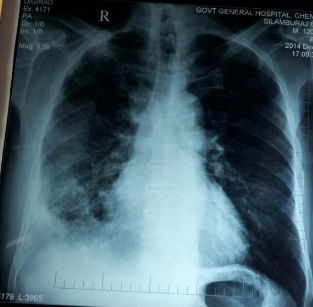
Reference

1. Bryan M.Burt, Joseph B Shrager.Benign and Malignant Neoplasm of the mediastinum In:Miicheal A.Grippi editor. Fishman’s pulmonary diseases and Disorders 5th edn McGraw Hil;l Part10 Chapter 82 p.2675
2. Francis IR, Korobkin M.Pheochromocytoma. Radiol ClinNorth Am. 1996;34(6):1101-12.
3. Lin MW, Chang YL., Lee YC, Huang PM. Non-functionalparagangliom of the posterior mediastinum InteractCardiovasc Thoac Surg.2009;9(3):540-2.
4. Wald o, Shapira OM, Murar A, Izhar U.paragannglioma of the mediastinum: challenges in diagnosis and surgicalmanagement. j Cardiothorac Surg. 2010;5:19.
5. Jime ́nez C, Cote G, Arnold A, Gagel RF 2006 Review: Should patients.
6. with apparently sporadic pheochromocytomas or paragangliomas
7. be screened for hereditary syndromes? j Clin Endocrinol
8. Metab 91:2851–2858
9. Young WF jr:aragangliomas:clinical overview. Ann N Y Acad Sci 2006,1073:21-29.
10. Balcombe J, Torigian DA, Kim W Miller WT Jr: Cross- sectional imagine ofparagangliomas of the aorti body and other thoracic branchiomeric paraganglia. AJR Am J Roentgenol 2007, 188:1054-1058.
11.Wald et al., Paraganglioma of the mediastinum:  challengesin diagnosis and surgical management Journal of Cardiothoracic Surgery 2010,5:19
12. Mark W.Lingen Head and Neck Paraganglioma (Caroti dbody  tumour) In: ,editorRobbins &Cotran Pathologic  Basis of Disease, 9th ed P 741-742
13. Kliewer KE, Wen DR, Cancilla PA, Cochran AJ.Paragangliomas: assesment of proognosis byhistological, immunohistochemical andultrastructural tehniques. Hum Pathol . ;20:29-39,1989
14.Lamy AL, Fradet GJ, Luoma A, Nelems B: Anterior and middlemediastinum paraganglioma: complete resection is the treatment ofchoice  Ann Thorac Surg 1994, 57:249-252.
15.Zovato set al, Peptide Receptor Radionuclide Therapy (PRRT) with 77Lu-DOTATATE in Individual with Neck and Mediastinal paraganglioma (PGL) Horm Metab Res 2012;44: 411–414

803 views

Non Resolving Pneumonia – A Rare Differential

By | volume3-issue1 | No Comments

699 views

Monisha Anandhan et al.: Non Resolving Pneumonia – A Rare Differential

Case Report

Non Resolving Pneumonia – A Rare Differential

Monisha Anandhan1, Sridhar R2, Prabhu 3, Narasimhan.R4

1Post graduate–Department of Respiratory Medicine, Apollo Hospitals, Greams Road, Chennai, India
2Associate consultant Pulmonologist, Apollo Hospitals, Greams Road, Chennai, India
3Consultant Hematologist, Apollo Hospitals, Greams Road, Chennai, India
4Senior consultant Pulmonologist, Apollo Hospitals, Greams Road, Chennai, India

Abstract

Multiple myeloma(MM) is a malignant systemic hematological disease that arises from monoclonal plasma cells. It usually affects older patients and is characterized by the presence of monoclonal immunoproteins in the serum. They are rarely found in extramedullary sites. We present a case of an elderly female who came to us with Respiratory symptoms, myalgia and loose stools for 2 weeks, not responding to standard antibiotic therapy raising an alarm of non resolving pneumonia of non-infectious etiology. Further radiological and laboratory evaluations showed diffuse parenchymal infiltrate and plasmacytosis. Thus the diagnosis of Multiple myeloma was made.
Keywords: Non resolving pneumonia, Plasma cells, Multiple myeloma.

Corresponding Author: Dr. Monisha Anandhan, Department of Respiratory Medicine, Apollo Hospitals, Greams Road, Chennai,
India. Email: monishaanandhan49@gmail.com

Introduction
Multiple myeloma is a plasma cell dyscrasia with an incidence of 10% of all hematologic malignancies. It is characterized by proliferation of myeloma cell clones and abnormal protein production. Infections are the most common complications in multiple myeloma, followed by end-organ damage .MM characterized by renal insufficiency, hypercalcemia, anemia, or lytic bone lesions.(1)The incidence of extramedullary manifestation is less than 7% among which pleural and pulmonary parenchymal involvement are extremely rare which usually Journal of the Association of Pulmonologist of Tamil Nadu appear late in the course of the disease.

Multiple myeloma may produce varied patterns on chest radiographs including diffuse interstitial disease from alveolar septal amyloidosis, consolidation, masses mimicking as solid tumour metastasis and mediastinal lymphadenopathy.(2) The parenchymal causes of respiratory failure from multiple myeloma include alveolar septal amyloidosis, plasma cell infiltration of lung parenchyma, accumulation of alveolar paraproteins, and metastatic calcification of the alveolar walls and blood vessels.(3)

Patients with clinical diagnosis of community-acquired pneumonia who fail to respond to antibiotics should be worked for non resolving pneumonia. The term non resolving pneumonia or slowly resolving pneumonia have been used interchangeably .It is difficult to distinguish between pulmonary manifestations of multiple myeloma and pulmonary infections in multiple myeloma secondary to impaired host immunity.

How to cite this article: Monisha Anandhan, Sridhar R, Prabhu, Narasimhan.R, Non Resolving Pneumonia – A Rare Differential, JAPT 2020; 3(1):42-46

Case Report

A 65 year old, Hypertensive, Anaemic, morbidly obese lady, presented to us with the complaints of breathlessness, cough with expectoration, myalgia and loose stools for a duration of 2 weeks. On examination, the patient was tachypneic and Spo2 was 88% on room air and onauscultation bilateral crepitations was present. Laboratory investigations showed hemoglobin of 9.2 g/dl, total leukocyte count 22,254 cells/mm3, Kidney function tests of serum creatinine of 1.4 mg/dl and hypoproteinemia. Chest radiograph and computerized tomography (CT) of chest showed bilateral diffuse parenchymal infiltrates (Fig 1&2). Provisional diagnosis of community acquired pneumonia with sepsis and acute kidney injury was madeand started on IV antibiotics (Cefoperazone and sulbactamand azithromycin). Her sputum for bacterial culture, Xpert MTB and respiratory viral panel were negative and blood culture showed no growth. Her ECHO revealed LVH, mild dilatation of RA, RV, mild PAH, EF-60%. computerized tomography (CT) of chest done which revealed bilateral asymmetric patchy central and peripheral consolidation relatively sparing subpleural space predominantly in upper lobes with bilateral pleural effusion. Pleural fluid analysis shows transudative effusion and negative for malignant cells. Due to persistent kidney dysfunction, she was initiated on hemodialysis. Her liver enzymes and bilirubin were elevated, secondary to sepsis with persistent cholestasis. Repeat chest radiograph showed no improvement in opacities (Fig 3) and her impaired kidney function was persisting. In the view of non resolving pneumonia, vasculitis was suspected and ruled out. Repeat hemogram revealed fall in Hb and persistently elevated white cell counts with increased differential count of plasma cells for which Hematologist opinion obtained and bone marrow aspiration done which was found to be hypercellular marrow with trilineagehaematopoiesis with plasmacytosis (plasma cells – 48), suggestive of Multiple Myeloma (MM) IgG stage 3 (as per Durie-Salmon staging system) and was correlated with serum free light chain assay (Free Kappa -2875). Her serum calcium were within normal limits. She was started on Bortezomib and Dexamethasone. Her repeat computerized tomography (CT) of chest showed resolving parenchymal infilterates (Fig 4). Despite of resolving radiographic picture, she developed Sepsis –MODS and was eventually declared.

Figure 1 – Intial X-Ray

Figure 2 – Pre treatment CT

Figure 3 – Non Resolving

Figure 4 – Post Treatment CT

Discussion

Community acquired pneumonia usually responds to empirical antibiotic therapy and attains clinical stability within 72 hours, if not then consider it as non resolving pneumonia. The entity of “slow resolution” has been defined as failure of radiographic resolution by 50% in 2 weeks or failure of complete resolution by one month despite adequate antibiotic therapy.(4) The causes include incorrect diagnosis, impaired host defence, inappropriate antimicrobial therapy, super infection, delayed radiological recovery, presence of resistant organism, disease mimicking pneumonia like non infectious causes which includes vasculitis and malignancy.(5) Delay in diagnosis and treatment may lead to rise of mortality by 3-5%. In this case the presence of multi-organ involvement (kidney, liver, heart), prolonged symptoms and non resolving opacity in chest radiograph in the absence of any microbiological evidence for infectious etiology arouse the suspicion of noninfectious etiology for non resolving pneumonia.

Initial differential diagnosis was pulmonary renal syndrome due to vasculitis. It is characterized by diffuse alveolar haemorrhage (DAH) and glomerulonephritis. The majority of cases of pulmonary-renal syndrome are ANCA associated but was negative in our case. Non resolving pneumonia along with kidney involvement and peripheral blood smear showing plasma cells, arouse the suspicion of Multiple myeloma. Initially plasma cells were considered as false positive due to infection and acute kidney injury and was treated as secondary to sepsis.

The plasma cells are terminally differentiated B cells which accounts for less than 3% in bone marrow. Plasma cell dyscrasia is neoplasm due to the expansion of a single clone of immunoglobulin secreting plasma cells. The common variants includes Monoclonal gammopathy of undetermined significance (62 %) and Multiple myeloma (18%)

The Multiple Myeloma represents 1% of neoplastic diseases and 13% of hematologic cancers. The risk of infection is 0.8% to 2.2% in patients with Multiple Myeloma mainly due to polyclonal hypogammaglobulinemia and is the leading cause of death often associated with bacterial infections of the lung.(6) The clinical manifestation of multiple myeloma includes anemia with a drop in hemoglobin of at least 2 g/dl compared to the normal value, or a hemoglobin below 10 g/dl, hypercalcemia (serum calcium >1 mg/dL than the ULN or >11 mg/dL), osteolytic lesions, renal failure and hyperviscosity syndrome.

Extramedullary myeloma (EMM) is plasma cell tumor arising outside the bone marrow. The most common primary site includes upper airway passage and oropharynx, rarely lymph nodes, liver, spleen and lungs. (7) In one large case series of 869 cases, there was no mention of lung involvement. Pulmonary presentation includes pulmonary infilterates, nodules, mass lesion, pleural effusion and thickening (7).

EMM is present in 6%–8% of patients at the time of myeloma diagnosis, and the incidence increases over the duration of the disease with 10%–30% of myeloma patients eventually presenting with EMM. The common complications include amyloid deposits causing damage to the affected organs. Pleural effusion is very rare, and occurs in <1% The most common cause of pleural effusions in patients with myeloma is congestive heart failure.(8) The pleural effusion develops at an average of 12 months after the diagnosis. In her case pleural fluid was tarnsudative and cytology was negative.

Diagnostic criteria for myeloma are A) ≥10% monoclonal plasma cells in the marrow or biopsy-proven plasmacytoma, B) Monoclonal protein in the serum and/or urine and C)Myeloma-related dysfunction of at least one organ system .(9) In her case she met with all these criteria, a diagnosis of Multiple Myeloma(MM) IgG stage 3 was made.

Definitive therapy is required when the patient is symptomatic or when organ dysfunction is present or impending. The initial treatment with MM varies depending on whether patients are eligibile or not to pursue autologous hematopoietic stem cell transplantation (HCT). All patients should be evaluated for eligibility criteria for autologous HCT. She did not meet the criteria based on her performance score and comorbidity and hence treated with aggressive chemotherapy (Bortezomib and dexamethasone). Combining bortezomib with other agents that are active in myeloma, such as corticosteroids, thalidomide, and low-dosage melphalan, increases its efficacy. The main adverse effects are thrombocytopenia, gastrointestinal disturbances, and peripheral neuropathy. Bortezomib, reversible proteasome inhibitor particularly effective in patients with light-chain disease causes rapid cytoreduction and its common side effects were nausea and peripheral neuropathies (35%). While her repeat imaging (CT-Chest) showed resolving opacities, she developed Septicemia and MODS (Multiorgan dysfunction syndrome). She was intubated in view of Respiratory distress and initiated on Renal Replacement Therapy. Unfortunately her body failed to respond to the treatment regimens and she was declared a week later.

Conclusions

Pulmonary parenchyma in multiple myeloma as a site of extramedullary involvement is rare and should be considered in patients who are presenting as non resolving pneumonia in selected situations. Peripheral smear and bone marrow biopsy are essential for diagnosis. Early intervention is paramount for prevention of morbidity and mortality in these patients. Since this can be treated with chemotherapy and stem cell transplantation if diagnosed earlier.

References

1. Kyle RA, Rajkumar SV. Multiple myeloma [published correction appears in N Engl J Med. 2005 Mar 17;352(11):1163]. N Engl J Med. 2004;351(18):1860-1873
2. Abhishek K, Ejazi MA, Hashim Z, Chaudhary R, Niharika K. Multiple myeloma with different thoracic manifestations: Case series. Indian J Respir Care 2018;7:108-12
3. Ramnik K, Duggal RK, Ramachandran KA. Multiple myeloma with extramedullary dissemination in the lung. JIACM 2002;3:93-5
4. Rome L, Murali G, Lippmann M. Nonresolving pneumonia and mimics of pneumonia. Med Clin North Am 2001;85:1511-30.
5. Prasad R, Verma SK, Sodhi R. Multiple myeloma with lung plasmacytoma. Lung India 2011;28:136-8
6. Alves dos Santos JW, Torres A, Michel GT, et al. Noninfectious and unusual infectious mimics of community-acquired pneumonia. Respir Med. 2004;98(6):488‐494.
7. Joseph G, Pandit M, Korfhage L. Primary pulmonary plasmacytoma. Cancer. 1993;71(3):721-724.
8. Rodríguez JN, Pereira A, Martínez JC, Conde J, Pujol E. Pleural effusion in multiple myeloma. Chest. 1994;105(2):622-624
9. Rajkumar SV, Kyle RA. Multiple myeloma: diagnosis and treatment. Mayo Clin Proc. 2005;80(10):1371-1382.
10. O’Sullivan P, Müller NL. Pulmonary and nodal multiple myeloma mimicking lymphoma. Br J Radiol. 2006;79(943):e25-e27.
11. Marmor DB, Farber JL, Gottlieb JE. Acute respiratory distress syndrome due to pulmonary involvement by neoplastic plasma cells in multiple myeloma. Thorax. 2006;61(5):455-456.

400 views

A Case Report of Extramedullary Plasmacytoma – Presenting as a Chest Wall Tumour

By | volume3-issue1 | No Comments

699 views

S. Priya et al.: A Case Report of Extramedullary Plasmacytoma – Presenting as a Chest Wall Tumour

Case Report

A Case Report of Extramedullary Plasmacytoma – Presenting as a Chest Wall Tumour

S.Priya1, Murugan2, Mahilmaran 2

1Department of Pulmonology, Vellore Medical College, Vellore
2Institute of Thoracic Medicine, Madras Medical College, Chennai.

Abstract

Plasmacytoma refers to a malignant plasma cell tumor growing within soft tissue or within the axial skeleton. Plasmacytoma accounts for six percent of all primary chest wall tumours. A solitary extramedullary plasmacytoma is reported three percent of plasma cell disorders. A 65 year male patient presented with complaint of swelling in the back of right side of the chest noticed for one month, pain over the swelling and dry cough for two weeks. Physical examination revealed a diffuse, lobulated swelling in the back of right side of chest which is firm to hard in consistency. The chest radiograph showed a large extra-pulmonary opacity with well-defined medial margin and lateral margin merging with chest wall on right side and blunting of the right costophrenic angle. The histopathological examination of tru-cut biopsy section showed fragments of fibrocartilagenous stroma infiltrated by mononuclear cells with round to oval nucleus and eosinophilic cytoplasm and some foci showed plasmocytoid features with the intervening stroma shows proliferating capillaries thereby features suggestive of plasmacytoma. The specimen is positive for CD 138 by the immunohistochemistry analysis which is the hallmark for diagnosing plasmacytoma.
Keywords: Extramedullary Plasmacytoma, Tru-cut biopsy, Immunohistochemistry..

Corresponding Author: Dr. S. Priya, Assistant Professor, Department of Pulmonology, Vellore Medical College, Vellore

Introduction
A plasmacytoma is a discrete, solitary mass of neoplastic monoclonal plasma cells in either bone or soft tissue (extramedullary). Plasmacytoma accounts for six percent of all primary chest wall tumours. There are three distinct groups of plasmacytoma defined by the International Myeloma Working Group1:

Solitary plasmacytoma of bone (SPB), Extramedullary plasmacytoma (EMP) and Multiple plasmacytoma that are either primary or recurrent. A solitary extramedullary plasmacytoma is reported three percent of plasma cell disorders and it presents without bony or systemic involvement.

In the United States, the incidence is approximately 0.15 cases/100,000 person-years accounting for approximately 450 new cases per

How to cite this article: S.Priya, Murugan, Mahilmaran, A Case Report of Extramedullary Plasmacytoma – Presenting as a Chest Wall Tumour, JAPT 2020; 3(1):47-52

year. The incidence is highest in Blacks and lowest in Asians and Pacific Islanders. Men are diagnosed twice as frequently as women. The median age at diagnosis is 55 to 65 years, which compares with a median age at diagnosis of 71 years for patients with multiple myeloma (MM) 2,3,4. SPB has been reported in patients as young as 15 years 4,5,6. Although an increased risk of plasma cell dyscrasia has been reported in first- degree relatives of patients with monoclonal gammopathy of undetermined significance and patients with multiple myeloma, there are no data on familial predisposition in solitary plasmacytoma.
The diagnostic criteria for solitary bone plasmacytoma (SBP) with the use of magnetic resonance imaging (MRI), flow cytometry, and polymerase chain reaction (PCR) are as follows, single area of bone destruction due to clonal plasma cells, bone marrow plasma cell infiltration not exceeding 5% of all nucleated cells, absence of osteolytic bone lesions or other tissue involvement (no evidence of myeloma), absence of anemia, hypercalcemia, or renal impairment attributable to myeloma, low concentration of serum or urine monoclonal protein and preserved levels of uninvolved immunoglobulins.
The diagnostic criteria for extramedullary plasmacytoma (EMP) are as follows ,tissue biopsy showing monoclonal plasma cell histology, bone marrow plasma cell infiltration not exceeding 5% of all nucleated cells, absence of osteolytic bone lesions or other tissue involvement (no evidence of myeloma),absence of hypercalcemia or renal failure and a low serum M protein concentration.

Case Report

A 65 year male patient presented with complaint of swelling in the back of right side of the chest one month and pain in the swelling and dry cough for past two weeks. The painless swelling is noticed by the patient before one month which is insidious in onset and slowly progressive in size. The dull aching pain started later over the swelling brought the patient to seek medical help. The patient complained of shortness of breath and wheeze. He denied trauma to the chest wall. He was a former smoker with pack years of 20, consumes alcohol daily since 20 yrs of age and betal nut chewer. Physical examination revealed a diffuse, lobulated swelling in the back of right side of the chest which was firm to hard in consistency, not mobile, not tender, no warmth and no dilated veins over the swelling, not fixed to the skin extending from inferior angle of scapula to posterior right costal margin approximately 20 x10 cm in size. On auscultation breath sound decreased in intensity in right infrascapular and infra axillary areas. The biochemical markers are normal. The ultrasonogram of the chest showed right pleural effusion and diagnostic thoracentesis proved to be exudative effusion The chest radiograph showed a large extra- pulmonary opacity with well-defined medial margin and lateral margin merging with chest wall on right side and blunting of the right costo phrenic angle. The contrast enhanced chest computed tomogram feature was a large homogenous enhancing soft tissue mass lesion in right prevertebral region with large exophytic chest wall component and dural component causing widening right neural foramina in fifth and sixth thoracic vertebra with mass effect on posterior aspect of right lung with right pleural effusion. Fine needle aspiration cytology reported by pathologist as cellular smear showing predominantly singly dispersed round to oval cells with moderate amount of cytoplasm and eccentrically placed nuclei, numerous binucleated, multinucleated cells with numerous mitotic figure suggestive of Plasmacytoma. The histopathological examination of tru-cut biopsy section showed fragments of fibrocartilagenous stroma infiltrated by mononuclear cells with round to oval nucleus and eosinophilic cytoplasm and some foci showed plasmocytoid features with the intervening stroma shows proliferating capillaries thereby features suggestive of Plasmacytoma. The specimen is positive for CD 138 by the immunohistochemistry analysis which is the hallmark for diagnosing  Plasmacytoma.

Picture of the case showing swelling in the back

Figure 2 – Chest radiograph of the case

Figure 3 – Chest Computed tomograph of the case


Figure 4 – Fine needle aspiration cytology section showing round to ovel cells with moderate amount of cytoplasm and eccentrically placed nuclei

Figure 5 – Sectioo to tru cut biopsy specimen fibrocartilagenous stroma infiltrated by mononuclear cells with round to oval nucleus and eosinophilic cytoplasm


Bone marrow biopsy section showed bony trabaculae with intervening normocellular marrow showing myeloid erthroid precursors and adequate megakaryocte. No evidence of multiple myeloma in the material, Urine Bence jones protien was negative. The urine protien electrophoresis showed M – band.The serum protein electrophoresis revealed paraprotein band which is present in the gamma region.Thus the solitary extramedullary plasmacytoma is diagnosed in this case.

Treatment

Radiotherapy is the main choice of treatment for both solitary plasmacytoma of bone and extramedullary plasmacytoma, and local control rates of >80% can be achieved. This form of treatment can be used with curative intent because plasmacytoma is a radiosensitive tumor. Surgery is an option for extramedullary plasmacytoma, but for cosmetic reasons it is generally used when the lesion is not present within the head and neck region. NCCN guidelines – Primary solitary plasmacytoma treatment for Osseousis local radiotherapy, for extraosseous radiotherapy followed by surgery, adjuvant chemotherapy may be considered for patients with tumours larger than 5 cm. In this patient, proteosome inhibitor Inj. Bortezomib and T.Lenalidomide,an anti-tumor drug that has been shown to interact with ubiquitin E3 ligase cereblon enzyme inside the tumor cell was given, patient improved symptomatically with reduction in size of tumor with two cycles of chemotherapy and and its mass effect on the right lung is revealed.

Figure 6: Post Chemotherapy

Figure 7: Post chemotherapy – Chest Radiography

Discussion

The diagnosis of plasmacytoma uses a diverse range of interdisciplinary techniques including serum protein electrophoresis, bone marrow biopsy, urine analysis for Bence Jones protein and complete blood count, plain film radiography, MRI and PET-CT.8,9

Serum protein electrophoresis separates the proteins in the serum to analysis the antibodies. Normal blood serum contains a range of antibodies and are said to be polyclonal, whereas serum from a person with plasmacytoma may show a monoclonal spike. This is due to an outgrowth of a single type of plasma cell that forms the plasmacytoma and produces a single type of antibody. The plasma cells are said to be monoclonal and the excessively produced antibody is known as monoclonal protein or paraprotein.11 Paraproteins are present in 60% of solitary bone plasmacytoma and less than 25% of extramedullary plasmacytoma.

Bone marrow biopsies are performed to ensure the disease is localised; and in solitary plasmacytoma of bone or extramedullary plasmacytoma there will not be an increase of monoclonal plasma cells which is similar in this case as there is no evident of multiple myeloma
in the bone marrow biopsy. Tissue biopsies of solitary plasmacytoma of bone and extramedullary plasmacytoma are used to assess the phenotype of the plasma cells. Histological analyses can be performed on these biopsies to see what Cluster of differentiation (CD) markers are present and to assess monoclonality of the cells. CD markers can aid in the distinction of extramedullary plasmacytoma from lymphomas. In our patient, CD 138 is positive which is the pathognomic for plasmacytoma.

Skeletal surveys are used to ensure there are no other primary tumors within the axial skeleton. MRI can be used to assess tumor status and may be advantageous in detecting primary tumors that are not detected by plain film radiography. PET-CT may also be beneficial in detecting extramedullary tumours in individuals diagnosed with SPB. CT imaging may be better than plain film radiography for assessing bone damage.8,9

An important distinction to be made is that a true plasmacytoma is present and not a systemic plasma cell disorder, such as multiple myeloma. The difference between plasmacytoma and multiple myeloma is that plasmacytoma lacks increased blood calcium, renal insufficiency, anemia and multiple bone lesions (collectively termed CRAB).The reported patients’ blood calcium and renal function are normal as well as blood hemoglobin level of 11.5 gm%.

As mentioned in the reference articles11,12, for the distinction from solitary bone plasmacytomain extramedullary plasmacytoma there is the presence of only one lesion (either in bone or soft tissue), normal bone marrow (<5% plasma cells), normal skeletal survey, absent or low paraprotein and no end organ damage which was found in the present case. Most cases of solitary plasmacytoma of bone progress to multiple myeloma within 2–4 years of diagnosis, but the overall median survival for solitary plasmacytoma of bone is 7–12 years. 30– 50% of extramedullary plasmacytoma cases progress to multiple myeloma with a median time of 1.5–2.5 years. 15–45% of SPB and 50–65% of extramedullary plasmacytoma are disease free after 10 years.12

Conclusions

In this case bone marrow biopsy and immunochemistry report of CD 138 positive ruled out the diagnosis of multiple myeloma and confirmed the diagnosis of extramedullary
plasmacytoma, hence patient treated with chemotherapy and outcome is satisfactory. Any patient in the median age group of 55 presenting with soft tissue swelling with no skeletal involvement, early suspicious and diagnosis of benign form of tumor can be treated with neo- adjuvant chemotherapy.

References

1. International Myeloma Working Group (June 2003). “Criteria for the classification of monoclonal gammopathies, multiple myeloma and related disorders: a report of the International Myeloma Working Group”. Br. J. Haematol. 121 (5): 749–57.
2. Dores GM, Landgren O, McGlynn KA, et al. Plasmacytoma of bone, extramedullary plasmacytoma, and multiple myeloma: incidence and survival in the United States, 1992-2004. Br J Haematol 2009; 144:86.
3. Shih LY, Dunn P, Leung WM, et al. Localised plasmacytomas in Taiwan: comparison between extramedullary plasmacytoma and solitary plasmacytoma of bone. Br J Cancer 1995; 71:128.
4. Frassica DA, Frassica FJ, Schray MF, et al. Solitary plasmacytoma of bone: Mayo Clinic experience. Int J Radiat Oncol Biol Phys 1989; 16:43.
5. Pavithran K, Doval DC, Rao CR, et al. Pediatric solitary plasmacytoma. Acta Oncol 1997; 36:83.
6.Boos N, Goytan M, Fraser R, Aebi M.et al Solitary plasma- cell myeloma of the spine in an adolescent. Case report of an unusual presentation. J Bone Joint Surg Br 1997; 79:812.
7. Greenberg AJ, Rajkumar SV, Vachon CM.et al Familial monoclonal gammopathy of undetermined significance and multiple myeloma: epidemiology, risk factors, and biological characteristics. Blood 2012; 119:5359.
8. Kilciksiz S, Karakoyun-Celik O, Agaoglu FY, Haydaroglu A (2012).et al “A review for solitary plasmacytoma of bone and extramedullary plasmacytoma”. Scientific World Journal. 2012:895765.
9. Dimopoulos M, Terpos E, Comenzo RL, et al. (September 2009). “International myeloma working group consensus statement and guidelines regarding the current role of imaging techniques in the diagnosis and monitoring of multiple Myeloma”. Leukemia. 23 (9): 1545–56.
10. O’Connell TX, Horita TJ, Kasravi B (January 2005).et al “Understanding and interpreting serum protein electrophoresis”. Am Fam Physician. 71 (1): 105–12.
11. M. Hughes; R. Soutar; H. Lucraft; R. Owen; J. Bird et al. “Guidelines on the diagnosis and management of solitary plasmacytoma of bone, extramedullary plasmacytoma and multiple solitary plasmacytomas.
12. Weber DM (2005). “Solitary bone and extramedullary plasmacytoma”. Hematology Am Soc Hematol Educ Program: 373–6.

405 views

Radiology Quiz

By | volume3-issue1 | No Comments

699 views

T. Suresh Babu et al.: Radiology Quiz

Pictorial CME

Radiology Quiz

T.Suresh Babu1, Narasimhan R2

1Post Graduate, Department of Respiratory Medicine, Apollo Hospitals, Greams Road Chennai
2Senior Consultant, Department of Respiratory Medicine, Apollo Hospitals, Greams Road Chennai

Corresponding Author: Dr. Suresh Babu, Department of Respiratory Medicine, Apollo Main Hospitals, 21, Greams Lane, off
Greams Road, Chennai- 600 006, Tamil Nadu, India

This 58 years old lady who is a known case of Diabetic Nephropathy, Hypertension, came to OPD with the complaints of dry cough for 6 months, more in the night and hoarsness of voice. On auscultation breath sounds were reduced in left supraclavicular region. Chest x- ray,CT Chest and bronchoscopy images are as follows.
Guess the Diagnosis and sign?

How to cite this article: T.Suresh Babu, Narasimhan R, Radiology Quiz, JAPT 2020; 3(1):53-54

Atelectasis

Partial collapse or incomplete inflation of the lung. The terms “atelectasis”, “Loss of lung volume” and “collapse” are synonymously used.

Types of Atelectasis

  • Resorptive Atelectasis: When airways are obstructed there is no further ventilation to the lungs and beyond.
  • Relaxation Atelectasis: The lung is held close to the chest wall because of the negative pressure in the pleural space.
  • Adhesive Atelectasis- Occurs due to decreased production of pulmonary surfactant
  • Cicatricial Atelectasis- Occurs due to endobronchial lesion or mucus plug..
  • Round Atelectasis- Infolding of redundant pleura.

Major and Minor sign of lung collapse1,2

Major signs:

1. Crowding of pulmonary vessels
2. Displacement of fissures

Minor signs:

1. Elevation of Ipsilateral Diaphragm
2. Crowding of Ipsilateral ribs
3. Shift of mediastinum towards the side of collapse
4. Compensatory Hyperinflation of normal lung.
5. Hilar displacement towards the collapse.

Left Upperlobe Collapse

The above chest x-ray shows left para aortic hyperlucency and rib crowding. The above features suggestive of Luftsichel (Agerman word ; Luft Air and Sichel = Sickel) sign3. As there is no minor fissure onthe left, the appearance of the left upper lobe atelectasis is different from Right upper lobe atelectasis. The Lobe moves predominantly forward, pulling the expanded left lower lobe behind it. Except at the edges the lobe retains much of its original contact with anterior chest wall and mediastinum. Since the lobe thins as the fissure is pulled forward, the usual appearance on a frontal radiograph is a hazy density extending out from the left hilum, often reaching the lung apex, fading laterally and inferiorly. With increasing loss of volume the upper margin of aortic knob once again becomes visible because the superior segment of the lower lobe takes the place of the posterior segment of left upper lobe – a sign that has been called the “luftsichel sign”4. Ct scan chest was done for this patient, which showed left upper lobe collapse, followed by this he underwent bronchoscopy, which showed a mass lesion completely obstructing left upper lobe bronchus and cryobiopsy was done, HPE report showed leiomyoma.

References

1. Collins J, Stern EJ. Chest radiology, the essentials. Lippincott Williams & Wilkins. (2007)
2. Grainger RG, ADAM A, Dixon AK. Grainger & Allisons Diagnostic Radiology
3. Churchill Livingstone. W. Richard Webb, Charles B. Higgins. Thoracic Imaging. (2010)
4. Blankenbaker DG. The luftsichel sign. Radiology 1998; 208:319-320.


SITE LAUNCH

SITE LAUNCH