Prevalence of Prediabetes and Diabetes in 100 Newly Diagnosed Adult Asthmatics who Visited Pulmonology OPD of A Tertiary Care Hospital in South India – A Cross Sectional Study

By | volume2-issue3 | No Comments

Amal Johnson et al.: Prevalence of Prediabetes and Diabetes in 100 Newly Diagnosed Adult Asthmatics who Visited Pulmonology…

Prevalence of Prediabetes and Diabetes in 100 Newly Diagnosed Adult Asthmatics who Visited Pulmonology OPD of A Tertiary Care Hospital in South India – A Cross Sectional Study

Amal Johnson1, Mobeen Quadri2,Sumi M Sam3, Narasimhan R4

1Post Graduate, Department of Respiratory Medicine, Apollo Hospitals, Greams Road, Chennai, India
2Post Graduate, Department of Respiratory Medicine, Asram Medical College, Eluru, Andhra Pradhesh, India
3Physician Assistant, Department of Respiratory Medicine, Apollo Hospitals, Greams Road, Chennai, India
4Senior Consultant Pulmonologist, Apollo Hospitals, Greams Road, Chennai, India.

Abstract

Background: Diabetes mellitus is the most common endocrine disease and characterized by increased blood glucose levels. Diabetes has been associated with accelerated lung function decline, decrease in respiratory muscle strength altered bronchial reactivity. Methodology: This is a cross sectional study done in 100 consecutive patients >=18 years who visited Pulmonology OPD of Apollo Hospitals, Chennnai with respiratory symptoms and diagnosed to have asthma based on Spirometry according to GINA (2020) guidelines from March 2019 to May 2019. All patients with informed consent were subjected to serum HbA1C testing. Results: The results of HbA1C testing were as follows : 45 patients had HbA1C less than 5.6, 50 patients had HbA1C between5.6 to 6.4 which is classified as prediabetes and 5 patients had HbA1C greater than 6.4 which is classified as diabetes according to recent American diabetes association(ADA) guidelines. Conclusion:? There is a definite association of pre diabetes and diabetes with asthma and the drop in lung function correlates with the control of diabetes.

Keywords: Diabetes, Asthma, HbA1c, Prevalence.

Corresponding Author: Dr. Narasimhan R, Senior Consultant Department of Respiratory Medicine. Email:drrnarasimhan@gmail.com

How to cite this article: Amal Johnson, Mobeen Quadri, Sumi M Sam, Narasimhan R, Prevalence of Prediabetes and Diabetes in 100 Newly Diagnosed Adult Asthmatics who Visited Pulmonology OPD of A Tertiary Care Hospital in South India – A Cross Sectional Study, JAPT 2019; 2(3):94-99

Introduction

Diabetes mellitus (DM) comprises a group of metabolic disorders that share the common feature of hyperglycemia. DM is currently classified on the basis of the pathogenic process that leads to hyperglycemia. Type 1 DM is characterized by insulin deficiency, whereas type 2 DM is a heterogeneous group of disorders characterized by variable degrees of insulin resistance, impaired

insulin secretion, and excessive hepatic glucose production1,2. The global prevalence of DM is around 8.5% according to World Health Organization (2014)3. 366 million people will have DM by the year 2030. It is estimated that about In India, the prevalence is around 11.8%4. DM is a disease of significant morbidity and mortality in long term.

The diagnostic criteria for DM according to American Diabetes Association (ADA) include any one of the following5:

  • Fasting plasma glucose ≥7.0 mmol/L (≥126mg/dL)
  • Symptoms of diabetes plus a random blood glucose concentration ≥11.1 mmol/L (≥200mg/dL)
  • 2-h plasma glucose ≥11.1 mmol/L (≥200mg/dL) during a 75-g oral glucose tolerance test.
  • Hemoglobin A1c >6.5%

Prediabetes is an intermediate state of hyperglycemia with glycemic parameters above normal but below the diabetes threshold. It is defined as fasting blood glucose levels between 110mg/dl to 125mg/dl or HbA1C between 5.6 to 6.4%6.

HbA1C is glycosylated hemoglobin formed by non enzymatic addition of sugar to N-terminal of B chain which forms a Schiff base and converted into a Amadori product7. The use of HbA1C as a biomarker for monitoring the levels of glucose among diabetics was first proposed by Koenig et al8. It is a reliable measure of chronic glycemia and correlates well with the risk of long term diabetes complications. It is a convenient test, not requiring the patient to fast and uses only a single blood sample.

The metabolic, hormonal and microvascular disturbances frequently lead to kidney, retina, nerve and cardiovascular complications especially in uncontrolled DM. The damage to lungs resulting from chronic hyperglycemia has been studied recently and it occurs both in Type 1 and Type 2 DM.

The following complications are associated with DM1:

  • Increased lung infection
  • Pulmonary edema
  • Sleeping respiratory disorders
  • Reduced lung elasticity
  • Decreased CO diffusion
  • Altered bronchial reactivity
  • Increased risk of thromboembolism
  • Higher prevalence of pulmonary hypertension

The pathogenesis is related to the accumulation of advanced glycation end products in the alveolar basement membrane and oxidative stress9.
In patients with DM, there is histopathological evidence of thickened alveolar, epithelial and pulmonary capillary basal lamina10.
This study is an elementary attempt to estimate the prevalence of Prediabetes and Diabetes in newly diagnosed adult asthmatics.

Review of Literature

In the Framingham Heart study conducted on 3,254 participants, both FEV1 and FVC were lower in diabetics than in non-diabetics11.
In the COPD Gene study conducted with 10,129 participants, people with DM have reduced lung function after confounding for other known risk factors12.
In the Fremantle Diabetes Study done by Davis et al, there is an accelerated decline of FEV1 by 71ml/year and this decline related to poor glycemic control13.
In the Copenhagen City Heart Study conducted by Lange et al, FEV1 and FVC were consistently lower in diabetics, with a average reduction of 8% of the predicted value14.
The third national health and nutrition examination survey (NHANES III) conducted in 2005 over 1480 adults showed that there is a decrease of FEV1 and FVC in diabetics and the rate of decline associated with glycemic control15.
In meta analysis conducted by Borst et al on Diabetic pneumopathy, it is shown that lung function of diabetics is significantly impaired with 5% of predicted and correlates with glycemic state16..
In study entitled “Prevalence of bronchial asthma in patients with type 2 diabetes mellitus” by Themeli et al, 13.3% of the patients diagnosed to have diabetes had bronchial asthma17.
In an Australian retrospective, longitudinal cohort study done by Ehrlich et al, it is shown that the diabetics have increased risk of asthma (Hazard
ratio 1.08)18.
In cohort study conducted by Mueller et al entitled “ Asthma and the risk of type 2 diabetes in Singapore Chinese Health study” , asthma was associated with 31% increased risk of incident diabetes19.
In the sixth Korea National Health and Nutrition Examination Survey done by Kyung Hee Lee et al, the probability of asthma occurrence was 1.02 times higher20.

Objectives

To estimate the prevalence of Prediabetes and Diabetes in newly diagnosed adult asthmatics.

Materials and Methods

INCLUSION CRITERIA: Patients >=18 years diagnosed with Asthma based on spirometry according to GINA (2020) guidelines from March 2019 to May 2019 -FEV1 >=12% and 200ml.
EXCLUSION CRITERIA: Patients were excluded if age group <=18 or having other primary lung disease or already diagnosed to have diabetes.

Methodology :

This is a cross sectional study done in 100 consecutive patients >=18 years who visited Pulmonology OPD of Apollo Hospitals, Chennnai with respiratory symptoms and diagnosed to have asthma based on basis of variable symptoms (cough / wheeze / breathing difficulty / chest tightness) +/- reversible airflow limitation (spirometry – FEV1 > 12% and 200ml) according to GINA (2020) guidelines21 from March 2019 to May 2019. Demographic and clinical data collected
including Age, Gender, BMI, personal history of allergies (allergic rhinitis/atopic dermatitis), family history of asthma/allergies, smoking history, occupation history, history of respiratory infections and history of co morbidities and drug history. All patients with informed consent were subjected to
serum HbA1C testing.

Results

There were 70 males and 30 females in the study. The age range was between 19 to 58 and the average mean age was 35. The average BMI was 23 and 11 patients had BMI of >=30.
In patients with asthma, childhood history of allergy was present in 35% of patients and family history of asthma present in 35% of patients.
15 patients were smoking currently and 2 were former smokers. 3 patients had history of treated tuberculosis in the past.
In regard to co morbidities, 17 patients had systemic hypertension, 10 patients had underlying coronary artery disease, 4 patients had hypothyroidism and 3 patients had chronic kidney disease.
The results of HbA1C testing were as follows : 45 patients had HbA1C less than 5.6, 50 patients ad HbA1C between5.6 to 6.4 which is classified as prediabetes and 5 patients had HbA1C greater than 6.4 which is classified as diabetes according to recent American diabetes association(ADA) guidelines22.

The mean FEV1 of our patients was 64% and the mean bronchodilator reversibility was around 13%.
Out of the diagnosed as diabetes (HbA1C >- 6.5), 4 had moderate to very severe airflow obstruction in spirometry according to ATS/ERS 2005 classification23 , 24 out of 50 patients having prediabetes (HbA1C 5.6 -6.5) had moderate to very severe airflow obstruction in spirometry and 10 patients with HbA1C < 5.6 had moderate to very severe airflow obstruction in spirometry./p>

Discussion

There were 70 males and 30 females in the study and the sex ratio is 7:3. This is in discordant with the study results of Sofie et al24 where sex ratio of adult asthmatics diagnosed is 5:7. The average mean age was 35. This is in concordant with the study by Mirabelli et al.25 where the average age of
iagnosis in adult asthma is 38.

The average BMI was 23 kg/m2– Males with mean BMI of 24kg/m2 and Females with mean BMI of 23kg/m2. 11 patients had BMI of >=30 out of which 7 were females. This is in concordance with study done by Hjellvik et al.26 where the mean BMI in males was 26.2kg·m−2) than in females (24.7 kg·m−2), but severe obesity (BMI of ≥35kg kg·m−2) was more common in females than in males.

In patients with asthma, childhood history of asthma in 40% of patients, history of atopy in 70% of patients and family history of asthma present in
35% of patients. This is in concordance with the results of Moore et al.27.
But according to Siegal et al, there exists no relationship between atopy and development of diabetes mellitus and infact Cardwell et al. in their study have shown an inverse relationship between atopy and diabetes mellitus.

15 patients were smoking currently and 2 were former smokers. This is lesser than the prevalence of smoking in asthmatics is around 25% according to Parasuramalu et al.31 Also, asthmatic smokers are more prevalent for diabetes mellitus according to Song et al. due to exaggerated chronic inflammation.

In regard to comorbidities, 17 patients had systemic hypertension, 10 patients had underlying coronary artery disease, 4 patients had hypothyroidism and 3 patients had chronic kidney disease. This is in concordance with the study done by Xinming Su et al32. and Lee KH et al.20 where the most common co morbidity associated with asthma is hypertension. The possible pathway involved in development of hypertension in asthmatics is endothelin dysfunction and abnormal contraction of myosin II.

The results of HbA1C testing were as follows : 45 patients had HbA1C less than 5.6, 50 patients had HbA1C between 5.6 to 6.4 which is classified as prediabetes and 5 patients had HbA1C greater than 6.4 which is classified as diabetes according to recent American diabetes association(ADA) guidelines.
This prevalence of prediabetes among asthmatics in our study is 50% and is much more compared to our Indian prevalence of diabetes mellitus which is 14% according to Ramachandran et al33.

Also, the prevalence of diabetes among asthmatics in our study is 5% and is lesser than Indian prevalence of diabetes mellitus which is 11.8% according to Anjana RM et al.4

The prevalence of asthma and DM has been shown to range from 5% to 16% according to studies done by Adams et al34 and Caughey et al35 which correlates with our study.

The mean FEV1 of our patients was 64% and all patients had significant bronchodilator eversibility(>12% and 200ml). This is lesser than in study conducted by Sundeep Salvi et al.36 the mean FEV1 in diabetics was 78%.

The mean bronchodilator reversibility in diabetic asthmatics was 13% which is lesser than the mean of 21.5% in asthmatics according to Enrico Effler et al.37 which negatively correlates with asthma outcome.

We observed that 4 out of 5 patients had moderate to severe airflow obstruction who had HBA1C >=6.5%. and this is in concordance with study by Lee KH20 who showed that diabetics with higher HbA1C have increased risk of asthma occurence and poor asthma control.

According to Mueller et al38, possible mechanisms for increased risk of diabetes among asthmatics is

  • unregulated expression of
    proinflammatory pathways , including IL6, TNF-Alpha, adhesion molecules through activation of NF-KB
  • Hypoxia and intermittent hypoxia resulting in altered glucose metabolism via insulin dependent and insulin independent mechanisms
  • Exposure to glucocorticoids as treatment for diabetes affects glycemic control.

Limitation of the Study

    • To validate our findings in the study, we require large scale, multi-center randomized control trials.
    • Since this is a cross sectional study, we will follow up studies to establish the relationship between drop in lung function and control of diabetes.
    • HbA1C is usually measured with high performance liquid chromatography method (HPLC) and has variable results in patients with abnormal Red cell
      urnover like anaemia, Hb variants, immunosuppression, alcoholism and pregnant women39. Also, it has low positive predictive value in diagnosis of prediabetes and will need OGTT for confirmation of diagnosis40.

Conclusion

There is a definite association of pre diabetes and diabetes with asthma and the severity of irflow obstruction correlates with the HBA1C levels.

Acknowledgement

We sincerely thank the management of Apollo Hospitals for the constant support in our pursuit of excellence.

References

  1. Almeida R et al. DIABETIC PNEUMOPATHY. Brazilian Journal of Medicine and Human Health. 2016; 4(1).
  2. Kasper D, Fauci A, Hauser S et al. Harrison’s Principles Of Internal Medicine. New York: McGraw Hill Education; 2015.
  3. Diabetes mellitus, fasting blood glucose concentration, and risk of vascular disease: a collaborative meta-analysis of 102 prospective studies. Emerging Risk Factors Collaboration.
  4. Anjana RM et al. Prevalence of diabetes and prediabetes in 15 states of India: results from the ICMR–INDIAB population-based cross-sectional study. Lancet 2017;5(8):585-596.
  5. American Diabetes Association. Classification and Diagnosis of Diabetes: Standards of Medical Care in Diabetes-2020. Diabetes Care. 2020 Jan. 43 (Suppl1):S14- S31.
  6. Nidhi Bansal. Prediabetes diagnosis and treatment: A review. World J Diabetes. 2015 Mar 15; 6(2): 296–303.
  7. Sherwani S, Khan H, Ekhzaimy A, Masood A, Sakharkar M. Significance of HbA1c Test in Diagnosis and Prognosis of Diabetic Patients. Biomark Insights.016; 11:BMI.S38440. doi:10.4137/bmi.s38440.
  8. Koenig RJ, Peterson CM et al. Correlation of glucose regulation and hemoglobin A1C. N Engl J Med.1976;295:417-20
  9. Pitocco D, Fuso L, Conte E et al. The Diabetic Lung – A New Target Organ?. The Review of Diabetic Studies. 2012;9(1):23-35.
  10. . Weynaud B, Jonckheere A et al. Diabetes mellitus induces a thickening of the pulmonary basal lamina. Respiration 1999.66(1):14-19
  11. Walter RE, Beiser A et al. Association between glycemic state and lung function: the Framingham Heart Study. Am J Respir Crit Care Med. 2003; 167:911-6.
  12. Kinney GL, Black-Shinn JL et al. Pulmonary function reduction in diabetes with and without chronic obstructive pulmonary disease. Diabetes care. 2014; 37:389-95.
  13. Davis WA, Knuiman M, Kendall P, Grange V, Davis TM. Glycemic exposure is associated with reduced pulmonary function in type 2 diabetes: the Fremantle Diabetes Study. Diabetes Care 2004. 27(3):752-757.
  14. Lange P, Parner J, Schnohr P, Jensen G. Copenhagen City Heart Study: longitudinal analysis of ventilatory capacity in diabetic and nondiabetic adults. Eur Respir J 2002. 20(6):1406-1412
  15. McKeever TM, Weston PJ et al. Lung function and glucose metabolism: an analysis of data from the Third National Health and Nutrition Examination Survey. Am J Epidemiol 2005.161(6):546-556.
  16. Van der Borst et al. Diabetic pneumopathy:Evidence from a Meta-Analysis. Am J Respir Crit Care Med 179;2009:A1452
  17. Themeli Y, Ibro M, Dyrmishi L, Klosi J. Prevalence of bronchial asthma in patients with type 2 diabetes mellitus. Endocrine Abstracts. 2014.
    doi:10.1530/endoabs.35.p355
  18. Ehrlich S, Quesenberry C, Van Den Eeden S, Shan J, Ferrara A. Patients Diagnosed With Diabetes Are at Increased Risk for Asthma, Chronic Obstructive Pulmonary Disease, Pulmonary Fibrosis, and Pneumonia but Not Lung Cancer. Diabetes Care. 2009;33(1):55-60. doi:10.2337/dc09- 0880
  19. Mueller NT, Koh WP et al. Asthma and the risk of type 2 diabetes in the Singapore Chinese Health Study. Diabetes Res Clin Pract 2013 Feb 99(2):191-199.
  20. Lee KH and Lee HS. Hypertensionh and diabetes mellitus as risk factors for asthma in Korean adults: the Sixth Korea National Health and Nutrition Examination Survey. Int. Health 2019;00:1-7 21. 2020 GINA Main Report – Global Initiative for Asthma – GINA.
  21. 2020 GINA Main Report – Global Initiative for Asthma -GINA.
  22. American Diabetes Association. Standards of Medical Care in Diabetes – 2020 Abridged for primary care providers. Clinical Diabetes 2020 Jan;38(1):10-38.
  23. Pellegrino et al. Interpretative strategies for lung function tests. European Respiratory Journal 2005 26: 948-968;
  24. Sofie Hansen et al. Gender differences in adult-onset asthma: results from the Swiss SAPALDIA cohort study. European Respiratory Journal 2015 46: 1011-1020
  25. . Mirabelli et al. Age at asthma onset and subsequent asthma outcomes among adults with active asthma. Respir Med. 2013 Dec; 107(12): 1829–1836.
  26. Hjellvik V et al. Body mass index as predictor for asthma: a cohort study of 118,723 males and females. European Respiratory Journal 2010 35: 1235-1242
  27. Perez MK et al. Metabolic asthma: Is there a link between obesity,diabetes and asthma?. Immunol Allergy Clin North Am. 2014 November; 34(4): 777–784.
  28. Moore et al. National Heart, Lung, and Blood Institute’s Severe Asthma Research Program. Am J Respir Crit Care Med. 2010 Feb 15; 181(4):315-23.
  29. Seigal S and Herzstein J. Atopy and diabetes mellitus. Journal of Allergy and Clinical immunology. 1954;25(1): 25-27
  30. Cardwell CR et al. A Meta-Analysis of the Association between Childhood Type 1 Diabetes and Atopic Disease. Diabetes Care 2003 Sep; 26(9): 2568-2574.
  31. Parasumaralu BG et al. Prevalence of bronchial asthma and its association with smoking habits among adult population in rural area. Indian J Public Health 2010; 54:165-8.
  32. Xinming Su et al. Prevalence of Comorbidities in Asthma and Nonasthma Patients: A Meta-analysis. Medicine (Baltimore).2016 May;95(22):e3459
  33. Ramachandran A, Snehalatha C, Kapur A, Vijay V, Mohan V, Das AK, et al. High prevalence of diabetes and impaired glucose tolerance in India: National Urban Diabetes Survey. Diabetologia. 2001;44:1094–101.
  34. Adams RJ, Wilson DH et al. Coexistent chronic conditions and asthma quality of life: a population-based study. Chest 2006;129(2):285-91.
  35. Caughey GE et al. Prevalence of comorbidity of chronic diseases in Australia. BMC public health. 2008;8(1):221
  36. Sundeep Salvi et al. Pulmonary Function Tests in Type 2 Diabetes Mellitus and Their Association With Glycemic Control and Duration of the Disease. Lung India 2013 Apr;30(2):108-12.
  37. Enrico Heffler et al. Bronchodilator response as a marker of poor asthma control. Respiratory Medicine 112(2016) 45-50.
  38. Mueller NT et al. Asthma and the risk of type 2 diabetes in the Singapore Chinese study. Diabetes Res Clin Pract 2013 February ; 99(2):192-199.
  39. CS L, TC A. HbA1c in the diagnosis and management of diabetes mellitus: an update. Diabetes Updates. 2020;6(1).
  40. Aidar Gosmanov, Jim Wan. ow Positive Predictive Value of Hemoglobin A1c for Diagnosis of Prediabetes in Clinical Practice.Am J Med Sci. 2014 Sep;348(3):191-4

Role of Rigid Thoracoscopy in Undiagnosed Pleural Effusion: A Prospective Study

By | volume2-issue3 | No Comments

A. Manimaran et al.: Role of Rigid Thoracoscopy in Undiagnosed Pleural Effusion: A Prospective Study

Role of Rigid Thoracoscopy in Undiagnosed Pleural Effusion:
A Prospective Study

A. Manimaran1, K.Anbananthan2, A. Nadhiya3, R. Ponnar3

1Senior Resident, Department of Thoracic Medicine, Thanjavur Medical College and Hospital, Thanjavur, Tamil Nadu, India,
2Associate Professor, Department of Thoracic Medicine, Thanjavur Medical College,
3Junior Resident, Department of Thoracic Medicine, Thanjavur Medical College, Tamil Nadu, India

Abstract

Background: Medical thoracoscopy is a minimally invasive, safe, and cost-effective procedure that allows complete visualization of pleural space, enabling diagnostic and therapeutic procedures such as pleural biopsy and talc insufflations. Conservative estimates suggest that 25% of patients seen in general pulmonologist’s practice involve pleura, of these 25% are unable to be attributed to a specific diagnosis, even after thoracentesis and closed pleural biopsy. Aim: The aim of this study was to evaluate the diagnostic yield of rigid thoracoscopy in undiagnosed pleural effusion. Methods: This prospective observational descriptive study was conducted in Thanjavur Medical College Hospital, Tamil Nadu, in the Department of Thoracic Medicine between July 2017 and December 2018. Rigid thoracoscopy equipment was used for investigation. 12 undiagnosed pleural effusion patients after thoracentesis who fulfilled inclusion and exclusion criteria were included in the study. Results: Of the 12 patients, six were male and six were female; seven had right-sided pleural effusion and five had left sided pleural effusion. Investigation reports were inconclusive except for suggesting an exudative effusion. Cytological examinations of pleural fluid were inconclusive in all the patients. After thoracoscopy, tuberculosis was diagnosed in five cases, metastases in three cases, mesothelioma in two cases, and inflammatory pathology in two cases. Conclusion: Thoracoscopy is a safe procedure with high diagnostic yield in undiagnosed exudative pleural effusion (EPE). A simple, minimally invasive low-cost investigation reduces the need for more invasive and much more expensive thoracotomy.Our study proved that rigid thoracoscopy remains the investigation of choice in all undiagnosed EPE for
accurate diagnosis and management.

Keywords: Undiagnosed pleural effusion, Thoracentesis, Diagnostic yield, HPE

Corresponding Author: A. Manimaran, Senior Resident, Department of Thoracic Medicine, Thanjavur Medical College and Hospital,
Thanjavur, Tamil Nadu, India

How to cite this article: A. Manimaran, K.Anbananthan, A. Nadhiya, R. Ponnar, Role of Rigid Thoracoscopy in Undiagnosed Pleural Effusion: A Prospective tudy, JAPT 2019; 2(3):100-104

Introduction

Medical thoracoscopy also known as pleuroscopy is an endoscopic evaluation of the

pleural space. It is a minimally invasive procedure that was first invented by Hans Christian Jacobeus in 1910, who is regarded as “Father of Thoracoscopy.” Jacobeus also published an early report on the use of thoracoscopy to localize and diagnose benign and malignant lesions of the

pleura and pulmonary parenchyma.1 Medical
thoracscopy is usually done for the cases of
undiagnosed exudative pleural effusion (EPE). Pleural fluid aspirate for biochemical, cytological, and microbiological analysis is the initial investigation to diagnose the etiology. Closed pleural biopsy may help in additional cases, but complications such as bleeding and pneumothorax are common. Even after these two procedures, the final diagnosis is not arrived in 25–40% of effusions. There are two fundamental techniques by which thoracoscopy is performed, single puncture technique and double puncture technique.2 For single puncture technique, a 9-mm working channel is used, and for double puncture technique, a 7-mm working channel is used. The single puncture technique is commonly used by the chest physician.2 Thoracic ultrasound before thoracoscopy improves pleural access and predicts fibrous septation.3

Both rigid and semi-rigid thoracoscopes remain valuable in the evaluations and management of pleural disease, but rigid thoracoscope allows excellent visualization and inspection of the thoracic cavity and permits adequate sized biopsy specimens to be obtained.

Study Design and Materials

This is a prospective observational descriptive study conducted in the Department of Thoracic Medicine, Thanjavur Medical College Hospital, a tertiary care teaching hospital in Tamil Nadu. This is the second government medical college in Tamil Nadu, where thoracoscopy is being done. We included 12 patients with undiagnosed EPE. The study was done over 18 months from July 2017 to December 2018.

Undiagnosed pleural effusion was defined as failure to achieve diagnosis by microbiological, biochemical, and cytological analysis of pleural fluid.

Medical thoracoscopy investigation was done on these patients under general anesthesia after verifying that they met the inclusion and exclusion criteria. It was carried out in the Department of Thoracic Medicine, Thanjavur Medical College Hospital.
The inclusion and exclusion criteria are as follows:

Inclusion Criteria

The following criteria were included in the study:

  • Undiagnosed EPE as per Light’s criteria.
  • Adenosine deaminase (ADA) levels must be
  • Cytology was negative for malignancy. No underlying definitive lung pathology causing pleural effusion such as malignancy

Exclusion Criteria

The following criteria were excluded from the study:

  • Transudative pleural effusion as per Light’s criteria.
  • Pleural fluid ADA >40 IU/lt.
  • Pleural fluid cytology positive for malignancy.
  • Smear positive for pulmonary tuberculosis. Patient with bleeding diathesis.

Results

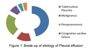
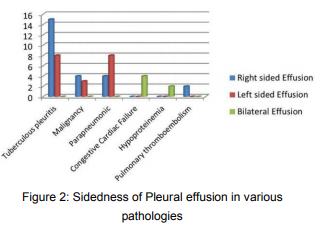
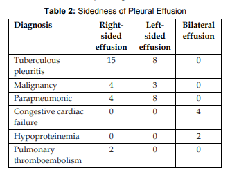
During inspection, the most common findings were adhesions, nodules, and sago grain appearance. Of the 12 patients, 6 were male and 6 were female. 7 had right- sided effusion and 5 had left-sided effusion. All the patients were in the age group of 41–65 years. Male-to-female ratio was 1:1,
the youngest was 41 years, and oldest was 65 years [Figures 1 and 2].

Nature and Color of the Pleural Effusion

All the hemorrhagic effusions turned out to be malignant, whereas in straw-colored effusions, five pateints had tuberculosis and two had inflammatory pathology.

Histopathology Results from Pleural Biopsy

Histopathology results confirmed malignancy in five cases, tuberculosis in five cases, while two cases were of inflammatory pathology.

Types of Malignant Lesion

Of five malignancies, three were metastatic adenocarcinomatous deposits and two cases were mesothelioma.


Medical thoracoscopy is a safe and valuable tool for the diagnosis of undiagnosed EPE, particularly for patients with a high probability of malignancy. The main indication in our study was A. Manimaran et al.: Role of Rigid Thoracoscopy in Undiagnosed Pleural Effusion: A Prospective Study 103 Journal of the Association of Pulmonologist of Tamil Nadu JAPT, Volume 2, Issue 3, Sep. – Dec. 2019 recurrent undiagnosed EPE, where etiology remains unexplained after initial and repeated cytological and biochemical analysis of pleural fluid.

Pleural effusion of undetermined etiology has been noted in all age groups. In our study, the age of the patients ranged from 41 to 65 years. In this
study, male and female are equal in number in contrast to many studies where male predominance is seen.4,5 The common thoracoscopic findings were adhesions, nodules, and sago grain appearance. All patients with nodules turned out to be malignant. This correlated with the study done by Helala et
al.5,6

As shown in Table 2 Tuberculosis and malignancy are the two most common HPE findings of pleural biopsy. As shown in Table 3 Only two types of malignant lesions were seen in our patients, metastatic adenocarcinomatous deposits followed by mesothelioma.

Pleural fluid cytology and closed pleural biopsy are two commonly applied procedures for the diagnosis of pleural effusion before thoracoscopy is performed. The diagnostic yield of cytology in malignant pleural effusion varies between 30% and 80%. Closed pleural biopsy increases the yield by about 10% and 40% in malignant and tuberculosis pleural effusions, respectively. However, bleeding and pneumothorax are common complications with closed pleural biopsy. Hence, its usage is minimal nowadays.

However, the diagnostic yield of thoracoscopy is about 83% in both malignant and tuberculous pleural effusions. Hence, thoracoscopy is an excellent diagnostic procedure as it provides direct visualization of the pleural surface which further increases the diagnostic yield.7

Maturu et al. had suggested that medical thoracoscopy is the procedure of choice in the evaluation of undiagnosed EPE, due to its higher success rate and an acceptable safety profile.8 Based on our findings, we concur with them on the same. Rigid thoracoscopy was found to be superior to semirigid thoracoscopy overall.9

Medical thoracoscopy can further progress with a wider adoption of the more interventional procedures and the improvement of equipment. The latter field includes minithoracoscopy.10

Conclusion

Medical thoracoscopy is a safe procedure with high diagnostic yield in undiagnosed EPE. A. Manimaran et al.: Role of Rigid Thoracoscopy in Undiagnosed Pleural Effusion: A Prospective Study 104 Journal of the Association of Pulmonologist of Tamil Nadu JAPT, Volume 2, Issue 3, Sep. – Dec. 2019 Malignant pleural effusion and tuberculosis are the common etiologies of undiagnosed exudative effusion.

Our study supports the notion that medical thoracoscopy remains the investigation of choice in all undiagnosed EPE for accurate diagnosis and management. However, the sample size is minimal, and hence, an elaborate study is needed to further strengthen our findings.

References

  1. Jacobaeus HC. The practical importance of thoracoscopy in surgery of the chest. Surg Gynaecol Obstet 1992;34:289-96.
  2. Loddenkemper R. Medical Thoracoscopy In: Light RW, Lee YCG, editors. Textbook of Pleural Diseases, 2nd ed. London, England: Hodder Arnold Publishers; 2008. p. 583-98.
  3. Medford AR, Agarwal S, Bennett JA, Free CM, Entwisle JJ.Thoracic ultrasound prior to medical thoracoscopy improves pleural access and predicts fibrous septation. Respirology 2010;15:804-8.
  4. Mootha VK, Agarwal R, Singh N, Aggarwal AN, Gupta D,Jindal SK, et al. Medical thoracoscopy for undiagnosed pleural effusions: Experience from a tertiary care hospital in North India. Indian J Chest Dis Allied Sci 2011;53:21-4.
  5. Helala LA, EI-Assal GM, Farghally AA, Abd EI Rady MM. Diagnostic yield of medical thoracoscopy in cases of undiagnosed pleural thoracoscopy in cases of undiagnosed pleural effusion in Kobri military hospital. Egy J Chest DiseTuber 2014;63:629-34.
  6. Prabhu VG, Narasimhan R, The role of pleuroscopy in undiagnosed exudative pleural effusion. Lung India 2012;29:128-30.
  7. Sodhi R. Sindhwani G. Nadia S, Kumar S, Jethani V, Khanduri S. A three year experience of medical thoracoscopy at a tertiary care center of Himalayan Region.J Cardiothorac Med 2016;4:397-402.
  8. Maturu VN, Dhooria S, Bal A, Singh N, Aggarwal AN, Gupta D, et al. Role of medical thoracoscopy and closedblind pleural biopsy in undiagnosed exudative pleural effusions: A single-center experienc of 348 patients. J Bronchology Interv Pulmonol 2015;22:121-9.
  9. Dhooria S, Singh N, Aggarwal AN, Gupta D, Agarwal R. A randomized trial comparing the diagnostic yield of rigid and semirigid thoracoscopy in undiagnosed pleural effusions. Respir Care 2014;59:756-64.
  10. Tassi G, Marchetti G. Minithoracoscopy: A less invasive approach to thoracoscopy. Chest 2003;124:1975-7.

Primary Tuberculosis of Upper Respiratory Tract – A Comprehensive Review Article

By | volume2-issue3 | No Comments

Amal Johnson et al.: Primary Tuberculosis of Upper Respiratory Tract – A Comprehensive Review Article

Primary Tuberculosis of Upper Respiratory Tract -A Comprehensive Review Article

1Post graduate, Department of Respiratory Medicine, Apollo Hospitals, Chennai
2Post graduate, Department of E.N.T., Stanley Medical College, Chennai
3Professor and Chief, Department of E.N.T., Stanley Medical College, Chennai

Abstract

Upper respiratory tract constitutes the first line of defense against all the inhalational agents. Laryngeal TB is the most common of all forms of upper respiratory tract TB (URT-TB). URT-TB is especially seen in immunocompromised patients such as presence of human immunodeficiency virus (HIV) infection, diabetes, smoking, alcoholism and use of immunosuppressive drugs. Granulation, Nodular or ulcerative lesions are seen on morphological examination. Endoscopic examination is required for mucosal lesions and granulation. Acid fast staining and histopathological examinations help in establishing the final diagnosis. Treatment includes standard anti-TB chemotherapy for 6 months..

Corresponding Author: Dr. Amal Johnson, Post graduate Department of Respiratory Medicine. Email: amal.johnson2210@gmail.

How to cite this article: Amal Johnson, Anto Sherly Sophia, V. Rajarajan, Primary Tuberculosis of Upper Respiratory Tract – A Comprehensive Review Article, JAPT 2019;2(3):105-116

Introduction

Tuberculosis of the upper airway is usually seen in conjunction with primary pulmonary tuberculosis.1 The continuous airflow and the smooth mucosal lining do not allow the mycobacteria to settle down in the respiratory tract, except the larynx5. Upper respiratory tract TB (URT-TB) is one of the rare forms of extra pulmonary TB (EPTB)15. Before treatment, patients with active pulmonary TB progressively deteriorated and often developed laryngeal, otological, nasal, paranasal, and pharyngeal involvement5.

Epidemiology:

The frequency of involvement may vary from all parts of the upper respiratory tract from the nose to the vocal cords and the larynx5. The most common site of TB in the head and neck involved the cervical lymph nodes and the nasopharynx11.Majority of cases of URT-TB have cervical lymphadenopathy2-9. Tubercular laryngitis is the most infectious form of disease16.

Tuberculous laryngitis manifest in isolation or in combination with tuberculosis of the epiglottis, pharynx, tonsils, or soft palate.13 while other patients had tuberculous otitis media, tuberculous tonsillitis, and tuberculous ulcerations of the tongue andpharynx14.

Common Symptoms and Signs of URT-TB

1. Nose – Nasal discharge, epistaxis, pain, nodule,
ulcer, septal perforation
2. Oral cavity – ulcer, localised swelling, tonsillar
infiltration, sore throat, dysphagia
3. Larynx – hoarseness, odynophagia, dysphagia.

Risk Factors Associated with URT-TB

  • HIV
  • Diabetes
  • Malignancies
  • Tobacco smoking
  • Drug abuse, alcoholism
  • Connective tissue disorders
  • Use of immunosuppressive drugs
  • Malnutrition
  • Poor living condition

Nasal Tuberculosis

Isolated cases of nasal tuberculosis have been reported in developing countries 4, 5, 10, 18–21

Clinical Features:

Patients with nasal TB commonly present with nasal obstruction and purulent rhinorrhea, Bloodstained discharge or frank epistaxis5,23,25. Lupus vulgaris, a slowly growing, indolent ulcerative lesion caused by Mycobacterium tuberculosis, may affect the nasal vestibule, the septum, and the alae. In few cases, lupus vulgaris with papulonecrotic TB is reported5,25,26. External deformity may result in about one-third of patients.5

Examination

    • Pallor of the nasal mucosa with multiple inute apple jelly nodules on diascopy.
    • Nasal septal ulceration and perforation of septal cartilage can occur2,21.
    • TB of nasal cavity can also present as polypoidal lesion27.
    • Sinonasal TB can invade the surrounding bones, causing osteomyelitis and abscess formation28.
    • Maxillary sinuses are commonly involved in nasal TB5.
    • Intracranial extension manifest as epilepsy and optic neuritis23,29
    • Intrasellar tuberculomas, also had involvement of sphenoid sinus 5,30

 width=

Confirmation of diagnosis is made on mycobacterial culture, histopathological examination, since the acid-fast bacilli (AFB) on smear examination may mimic Mycobacterium leprae. Standard anti-TB therapy is the treatment for Nasal TB5

 width=

Oral Cavity Tuberculosis:

TB is rarely involved in the oral cavity. Poor dental hygiene and mucosal injury contributed to the infection in the oral cavity. Oral lesions are the
result from the infected sputum being coughed out from concomitant pulmonary TB5, 38 . Due to hematogenous spread, The tongue is the most common site to be involved, almost any part of the tongue, such as the tip, the borders, dorsum, and base, may be involved. 5,39 .

 width=

Clinical Features

      • Single or multiple ulcers or Nodular lesions in outh manifest in oral cavity
      • Well circumscribed and painful irregular lesions mimicking malignancy.
      • Cervical lymphadenopathy also seen in oral cavity TB.2,40
      • A painful, deep, irregular ulcer on the dorsum of the tongue is classical oral mucosal lesion including the palate, lips, buccal mucosa and gingiva.19,39

 width=

A recent investigation reported positive oral cultures of M.tuberculosis from samples of saliva, caries lesions, and denture plaque collected from
TB patients, demonstrating the possibility of oral infectivity of these patients41,43. The detection rates from these oral sites using Polymerase chain reaction were higher (between 89% and 100% detection rates) than traditional culture methods (0% to 17%)42,43.

Tuberculosis of Pharynx:

TB of pharynx may present as ulcerative of lupus vulgaris type or secondary to pulmonary involvement (so-called miliary TB of the pharynx).5

Nasopharyngeal TB:

    • The nasopharynx is the most common site of pharyngeal involvement50.
    • Cervical lymphadenopathy was the most common presentation (58%) nasal obstruction (10%) tinnitus, Hearing loss, otalgia, rhinorrhea, and nasal twang of the voice,51snoring2.

Direct examination of the nasopharynx revealed a combination of mass and irregularity, granulation

      • On MRI, Two types of pattern of involvement in nasopharynxare seen: discrete polypoidal mass in the adenoids
      • Pattern of more diffuse soft tissue thickening of one or two walls of the nasopharynx52.
      • Extension outside the confines of the nasopharynx was not seen52.
      • Post radiation granulomatous inflammation in patients undergoing treatment for nasopharyngeal carcinoma should be suspected as occult tubercular infection and investigated thoroughly35.

Differential Diagnosis for Nasopharyngeal TB

      • Fungal infections
      • Malignancies
      • Following radiation therapy
      • Prolonged use of nasal spray
      • Wegeners granulomatosis
      • Midline granulomatous disease
      • Leprosy
      • Syphilis

Oropharyngeal Tuberculosis:

Oropharyngeal TB present with symptoms of sore throat, dysphagia, odynophagia53,54. TB retropharyngeal abscess may also present with dysphagia55.

Clinical Features of Oropharyngeal TB

    • Cervical lymphadenopathy
    • Cutaneous lupus vulgaris
    • Scrofuloderma
    • Local hyperemia and irregularity of mucosa
    • Erythematous papules
    • Swelling of the cheek are associated with oropharyngeal tuberculosis44.

Tonsillar Tuberculosis:

  • Tonsils is other site of involvement in Oropharyngeal TB which may occur in isolation or concomitant with pulmonary or laryngeal TB5.
  • Tonsillar TB was common in the ingestion unpasteurized milk contaminated with Mycobacterium bovis.

Clinical Features

    • Sore throat
    • cervical lymphadenopathy
    • dysphagia
    • ulceration
    • masses, and white patches are the features of Tonsillar TB56–58.
    • Pharyngeal TB can also spread to the middle ear through the eustachian tube59.
    • Multiple perforation of tympanic membrane, pale, painless profuse otorrhoea, Granulation and profound hearing loss, Preauricular lymph node enlargement and postauricular fistula are pathognomonic of tubercular otitis media.60
    • Physical examination includes unilateral tonsillar enlargement, ulcerations, and fibrosis of the tonsils. Incisional biopsy confirms the diagnosis based on histopathological findings and the identification of AFB 5.
    • Anti TB Chemotherapy is treatment for Tonsillar Tuberculosis.

Pale nodule in the oropharynx noticed incidentally during fiber optic bronchoscopy for mediastinal lymphadenopathy. Needle aspiration from the nodule revealed necrotizing granulomatous inflammation and multiple, pink stained AFB.5

Tuberculosis of Salivary Glands

    • TB of the salivary glands occurs as a result of infection of the oral cavity or secondary to pulmonary TB5,66.
    • Parotid involvement is the most common to be involved followed by submandibular glands67.
    • It could be acute or chronic parotidomegaly and diagnosis is obtained on histopathological evidence 61

Mode of transmission:

    • Tuberculous salivary gland infection is most common in older children and adults.
    • The disease is spread by close person to-person contact.
    • Primary salivary gland infection evolved from a focus in the tonsil or gingivobuccal sulcus before ascending to the glands by way of their ducts69.
    • Primary TB infections occur within the parotid gland then spread to the cervical nodes through the lymphatic drainage66
    • Other mechanisms include ascending lymphogenous spread from an infected cervical lymph node and hematogenous spread from a distant focus67.

    • Clinically, tuberculous salivary gland infection presents in two different forms.
    • The first is an acute inflammatory lesion with diffuse glandular edema.
    • A second is chronic, tumorous lesion is seen as a discrete slow-growing mass that mimics a neoplasm69.
    • The chest radiograph show evidence of healed granulomatous disease.

CT Features of TB Salivary Glands:

  • CT images of TB infection in the head and neck are described as having three patterns:
  • In early course of disease involvement of cervical lymphnodes with nonspecific homogenous enhancement.
  • In the second pattern, a nodal mass is apparent with central lucency and thick rims of enhancement and minimally effaced fascial planes.
  • The third pattern appears as fibrocalcified nodes, usually seen in patients previouslytreated for TB.72

Diagnosis:

  • Preoperative contrast-enhanced computed tomography (CT) show the presence of thickwalled rim-enhancing lesions with a central lucency suggests the diagnosis.
  • Tumors and other inflammatory processes show filling defects with or without thin walls.
  • Thick-walled round rim enhancing lesions with a central lucency are characteristic of TB.
  • Confirmation of the Diagnosis of most forms of extra laryngeal URT-TB requires biopsy5. On suspicion, diagnosis can also be made by fineneedle aspiration cytology61.
  • FNA biopsy is better than incisional biopsy due to lower risk of draining fistula in the former. The FNA sample reveal the characteristic cytologic features: granulomatous inflammation with caseous necrosis and epithelioid histiocytes.
  • Polymerase chain reaction (PCR) testing can help identify mycobacteria5,73 In addition, material may be sent for culture and acidfast smears; 5,74
  • When the diagnosis is uncertain or the lesion is resistant to medical therapy, complete surgical excision is both diagnostic and curative. 69

Treatment:

Treatment or all patients with pharyngeal and oral-cavity TB consists of anti-TB chemotherapy. The treatment response is generally favorable, and the prognosis is good62. Surgical intervention should be avoided63.

Laryngeal Tuberculosis

TB is the most common cause of granulomatous disease of the larynx. Tubercular laryngitis seems to be increasing recently6,7, 64–71. United States, Japan, and Spain, continue to point to the importance of the laryngeal TB5, 67, 72. Presently, laryngeal TB is reported in 1 to 2% of cases 1, 73.

Two cases of laryngeal TB were reported in renal transplant patients; both responded promptly to anti-TB therapy76

Rarely, patients on glucocorticoids can develop laryngeal TB. For example, a patient with Addison’s disease on glucocorticoids and another on inhaled steroid therapy are reported to have developed tubercular infection 77, 78. Laryngeal TB mimics a laryngeal carcinoma 79–81.

Clinical Features

    • Most patients seen today are without pulmonary symptoms or a history of pulmonary TB, and it is theorized that laryngeal disease is the result of
      hematogenous or lymphatic spread82,83.
    • The most common presenting symptom is hoarseness.82-84
    • The primary infection can involve any part of the larynx, predominantly involved the posterior larynx.
    • Other symptoms are dysphagia, odynophagia, cough, and weight loss.82,84
    • Despite the lack of pulmonary involvement, the purified protein derivative test result is usually positive85,86.

  • The lesions may be nodular, exophytic, or ulcerative,81,84-86 and because of their appearance, laryngeal TB is sometimes mistaken for squamous cell carcinoma (SCC).84.
  • Diagnosis of laryngeal TB is usually made by the combination of sputum culture,biopsy specimens that test positive for acidfast bacilli, and chest radiographs.84
  • Anti TB chemo therapy can alleviate symptoms and expedite the improvement in hoarseness89.
  • The true and false vocal folds were the most commonly affected sites.
  • Of these 60 patients, 28 (47%) had active pulmonary disease; 20 of the 60 (33%) had inactive pulmonary TB, and 9 (15%) had isolated laryngeal TB. Lim and colleagues made special note of the focal, atypical, and unilateral laryngeal findings in patients without active pulmonary disease.

Endoscopic features

    • Laryngeal edema and granulomatous involvement of the laryngeal mucosa present in laryngeal TB.
    • Granulation tissue at the level of the glottis, subglottic stenosis,
    • vocal cord paralysis secondary to mediastinal lymphadenopathy present with Upper airway obstruction.85.

  • Mucosal inflammation, hyperemia, mucosal edema,
  • Granulomatousmucosa, Mucosal ulcers ,
  • Localized swelling, abscess
  • Restricted movements of vocal cords, swelling/mass,
  • Polypoidal growth are seen in endoscopic appearance seen in Laryngeal Isolated involvement of the epiglottic, supraglottic, or subglottic region also involved 85–87.
  • A lateral X- ray of the neck and CT can help in differentiating TB from malignancy 90.
  • CT scan may show bilateral involvement, thickening of the free margin of the epiglottis, and preservation of the pre-epiglottic and paralaryngeal spaces even in the presence of extensive mucosal involvement in Laryngeal TB (90). Cartilage destruction is more common in malignancies and also seen in TB larynx 92.
  • Sputum microscopy is positive for 20% of patients with laryngeal TB.
  • Histopathological examination is required for a definite diagnosis.

The differential diagnosis of TB Larynx are

    • Bacterial and fungal infections,
    • granulomatosis with polyangiitis (Wegener’sgranulomatosis),
    • sarcoidosis, and
    • malignancies

Direct laryngoscopic examination with biopsy along with histopathological examination and culture provide the most conclusive evidence for diagnosis of Laryngeal TB. PCR-based analysis help to differentiate from other species of mycobacteria 36, 88.

Treatment and outcome:

The laryngeal lesions of TB respond well to standard anti-TB regimens, within weeks. The larynx is reported to return to its normal appearance in 18 weeks on average 73. Voice outcomes improve after anti-TB treatment in most patients 94. Vocal cord immobility due to fibrosis and adhesion may produce permanent hoarseness in few patients 95.

The standard treatment consists of four primary drugs (rifampin, isoniazid, pyrazinamide, and ethambutol) given together during an intensive phase of 2 months, followed by a maintenance phase of 2 or 3 drugs for 4 months. Surgical intervention such as tracheostomy, partial or complete laryngectomy, or laryngotracheoplasty may be required for some patients with abscess formation and progressive disease unresponsive to medical therapy.

In conclusion, TB should be kept in the differential diagnosis of upper airway diseases and/or cervical lymphadenopathy whenever a patient presents with hoarseness of voice/ obstructive symptoms ulcerative or granulomatous lesions, and failure of response to therapy for more common lesions.

It must be differentiated from malignancy from histopathological examination. Classic clinical features may not always be present. Early diagnosis and treatment are essential to prevent long-term complications of Tuberculosis.

References

  1. Chakravarti A, Pal S, Sahni JK (2008) Primary tuberculosis oftonsil and posterior oropharyngeal wall. Indian J Tuberc 55(1):48–50
  2. Menon K, Bem C, Gouldesbrough D, Strachan DR. 2007. A clinical review of 128 cases of head and neck tuberculosis presenting over a 10-year period in Bradford, UK. J Laryngol Otol 121:362–368.
  3. Prasad KC, Sreedharan S, Chakravarthy Y, Prasad SC. 2007. Tuberculosis in the head and neck: experience in India. J Laryngol Otol 121: 979–985.
  4. Akkara SA, Singhania A, Akkara AG, Shah A, Adalja M, Chauhan N.2014. A study of manifestations of extrapulmonary tuberculosis in the ENT region. Indian J Otolaryngol Head Neck Surg 66:46–50.
  5. Jindal SK, Jindal A, Agarwal R. 2016. Upper respiratory tract tuberculosis. Microbiol Spectrum 4(6):TNMI7-0009- 2016
  6. Das S, Das D, Bhuyan UT, Saikia N. 2016. Head and neck tuberculosis: scenario in a tertiary care hospital of northea stern India. J Clin Diagn Res 10:MC04–MC07.
  7. Bruzgielewicz A, Rzepakowska A, Osuch-Wójcikewicz E, Niemczyk K,Chmielewski R. 2014. Tuberculosis of the head and neck—epidemiological and clinical presentation. Arch Med Sci 10:1160–1166.
  8. Khuzwayo ZB, Naidu TK. 2014. Head and neck tuberculosis in KwaZulu-Natal, South Africa. J LaryngolOt ol 128:86–90.
  9. Al-Serhani AM. 2001. Mycobacterial infection of the head and neck: presentation and diagnosis. Laryngoscope 111:2012–2016.
  10. Tse GM, Ma TK, Chan AB, Ho FN, King AD, Fung KS, Ahuja AT.2003. Tuberculosis of the nasopharynx: a rare entity revisited. Laryngoscope 113:737–740.
  11. Srirompotong S, Yimtae K, Srirompotong S. 2003.Tuberculosis in the upper aerodigestive tract and human immunodeficiency virus coinfections. J Otolaryngol 32:230–233.
  12. Chiesa Estomba CM, Betances Reinoso FA, Rivera Schmitz T, OssaEcheverri CC, González Cortés MJ, Santidrian Hidalgo C. 2016. Head and neck tuberculosis: 6-year retrospective study. Acta Otorrinolaringol Esp 67:9–14.
  13. CASTLEMAN B, MCNEELY BU (editors): Case records of the Massachusetts General Hospital. N Engl J Med 279:423- 430,1968
  14. BULL TR: Tuberculosis of the larynx. Br Med J 2:991-992,1966
  15. Rohwedder JJ. 1974. Upper respiratory tract tuberculosis. Sixteen cases in a general hospital. Ann Intern Med 80:708–713.
  16. MR. 2004. Primary sinonasal tuberculosis in northwest Pakistan. J Coll Physicians Surg Pak 14:221–224. 17. Butt AA. 1997. Nasal tuberculosis in the 20th century. Am J Med Sci 313:332–335.
  17. Butt AA. 1997. Nasal tuberculosis in the 20th century. Am J Med Sci 313:332–335.
  18. Nawaz G, Khan Butt AA. 1997. Nasal tuberculosis in the 20th century. Am J Med Sci 313:332–335.
  19. MR. 2004. Primary sinonasal tuberculosis in northwest Pakistan. J Coll Physicians Surg Pak 14:221621.
  20. Butt AA. 1997. Nasal tuberculosis in the 20th century. Am J Med Sci 313:332–335.
  21. Nawaz G, Khan Butt –224
  22. Nalini B, Vinayak S. 2006. Tuberculosis in ear, nose, and throat practice: its presentation and diagnosis.
  23. Am J Otolaryngol 27:39–45.Masterson L, Srouji I,
  24. Kent R, Bath laboratory investigation. J Laryngol Otol 125:210–213.
  25. Sardana K, Goel K. 2014. Nasal septal ulceration. Clin Dermatol 32:817–826.
  26. Senol M, Ozcan A, Aydin A, Karincaoglu Y, Sasmaz S, Sener S. 2000. Disseminated lupus vulgaris and papulonecrotic tuberculid: case report. Pediatr Dermatol 17:133–135.
  27. Nayar RC, Al Kaabi J, Ghorpade K. 2004. Primary nasal tuberculosis: a case report. Ear Nose Throat J 83:188–191.
  28. Jha D, Deka RC, Sharma MC. 2002. Tuberculosis of the maxillary sinus manifesting as a facial abscess. Ear Nose Throat J 81:102–104.
  29. Das JC, Singh K, Sharma P, Singla R. 2003. Tuberculous osteomyelitis and optic neuritis. Ophthalmic Surg Lasers Imaging 34:409–412.
  30. Sharma MC, Arora R, Mahapatra AK, Sarat-Chandra P, Gaikwad SB,Sarkar C. 2000. Intrasellar tuberculoma—an enigmatic pituitary infection: a series of 18 cases. Clin Neurol Neurosurg 102:72–77.
  31. Thakur JS, Verma N, Mohindroo S, Azad RK, Mohindroo NK. 1 June 2016. Isolated tubercular hypoglossal nerve paralysis. Trop Doct
  32. Deepak D, Panjabi C, Gudwani S, Chaudhary N, Shah A.2001. Nasal septal perforation in a patient with allergic bronchopulmonary aspergillosis and rhinitis on long term corticosteroids. Asian Pac J Allergy Immunol 19:287–290.
  33. Lee CR, Yoo CI, Lee J, Kang SK. 2002. Nasal septum perforation of welders. Ind Health 40:286–289.
  34. Hughes RG, Drake-Lee A. 2001. Nasal manifestations of granulomatous disease. Hosp Med 62:417–421.
  35. Chan AB, Ma TK, Yu BK, King AD, Thakur JS, Verma N, Mohindroo S, Azad RK, Mohindroo NK. 1 June 2016.Isolated tubercular hypoglossal nerve paralysis. Trop Doct
  36. Deepak D, Panjabi C, Gudwani S, Chaudhary N, Shah A. 2001. Nasal septal perforation in a patient with allergic bronchopulmonary aspergillosis and rhinitis on long term corticosteroids. Asian Pac J Allergy Immunol 19:287–290.
  37. Lee CR, Yoo CI, Lee J, Kang SK. 2002. Nasal septum perforation of welders. Ind Health 40:286–289.
  38. Hughes RG, Drake-Lee A. 2001. Nasal manifestations of granulomatous disease. Hosp Med 62:417–421. Chan AB,Ma TK, Yu BK, King AD, Ho FN, Tse GM. 2004.
    Nasopharyngeal granulomatous inflammation and tuberculosis
  39. Nasopharyngeal granulomatous inflammation and tuberculosis complicating undifferentiated carcinoma. Otolaryngol Head Neck Surg 130:125– 130.
  40. Warndorff DK, Glynn JR, Fine PE, Jamil S, de Wit MY, MunthaliMM, Stoker NG, Klatser PR. 1996. Polymerase chain reaction of nasal swabs from tuberculosis patients and their contacts. Int J Lepr Other Mycobact Dis 64:404–408.
  41. Mycobacterium tuberculosis in oral cavity samples.Oral Microbiol Immunol 18:156–159,Kakisi OK,Kechagia AS, Kakisis IK, et al: Tuberculosis
    of the oral cavity: a systematic review. Eur J Oral Sci 118(2):103–109, 2010.
  42. Brook I: The bacteriology of salivary gland infections. Oral Maxillofac Surg Clin North Am 21(3):269–274, 2009.
  43. Eguchi J, Ishihara K, Watanabe A, et al:PCR method is essential for detecting Tuberculosis
  44. Morad NA. Tuberculous cervical lymphadenopathy; should antituberculous therapy be proceeded by histological proof? Tropical Doctor 2000; 30: 18–20.
  45. Aktoğu S, Eriş FN, Dinç ZA, Tibet G. 2000. Tuberculosis of the tongue secondary to pulmonary tuberculosis. Monaldi Arch Chest Dis 55:287–288.
  46. Iype EM, Ramdas K, Pandey M, Jayasree K, Thomas G, Sebastian P,Nair MK. 2001. Primary tuberculosis of the tongue: report of three cases. Br J Oral Maxillofac Surg 39:402–403.
  47. 47.Memon GA, Khushk IA. 2003. Primary tuberculosis of tongue. J Coll Physicians Surg Pak 13:604–605.
  48. 48.Dixit R, Sharma S, Nuwal P. 2008. Tuberculosis of oral cavity. Indian J Tuberc 55:51–53.
  49. Ilyas SE, Chen FF, Hodgson TA, Speight PM, Lacey CJ, Porter SR.2002. Labial tuberculosis: a unique cause of lip swelling complicating HIV infection. HIV Med 3:283–286.
  50. Kiliç A, Gül U, Gönül M, Soylu S, Cakmak SK, Demiriz M.2009. Orificial tuberculosis of the lip: a case report and review of the literature. Int J Dermatol 48:178–180.
  51. Ceballos-Salobreña A, Aguirre-Urizar JM, Bagan-Sebastian JV. 1996. Oral manifestations associated with human immunodeficiency virus infection in a Spanish population. J Oral Pathol Med 25:523–526
  52. Hale RG, Tucker DI. 2008. Head and neck manifestations of tuberculosis. Oral Maxillofac Surg Clin North Am 20:635–642.
  53. Miziara ID. 2005. Tuberculosis affecting the oral cavity in Brazilian HIV-infected patients. Oral Surg Oral Med Oral Pathol Oral Radiol Endod 100:179–182.
  54. Köktener A. 2001. Nasopharyngeal tuberculosis. Eur J Radiol 39:186–187.
  55. Aktan B, Selimoglu E, Uçüncü H, Sütbeyaz Y. 2002.Primary nasopharyngeal tuberculosis in a patient with the complaint of snoring. J Laryngol Otol 116:301–303.56.
  56. King AD, Ahuja AT, Tse GM, van Hasselt AC, Chan AB.2003. MR imaging features of nasopharyngeal tuberculosis:report of three cases and literature review. Am JNeuroradiol 24:279–282.
  57. Al-Serhani AM, Al-Mazrou K. 2001. Pharyngeal tuberculosis. Am J Otolaryngol 22:236-240
  58. Caylan R, Aydin K, Caylan R. 2002. Oropharyngeal tuberculosis causing severe odynophagia and dysphagia.Eur Arch Otorhinolaryngol 259:229–230.
  59. Christoforidou A, MetallidisS,Kollaras P,Agathangelidis A, NikolaidisP, Vital V, Markou K. 2012. Tuberculous retropharyngeal abscess as a cause of oropharyngeal dysphagia. Am J Otolaryngol 33:272–274
  60. Greenfield BJ, Selesnick SH, Fisher L, et al:Aural tuberculosis. Am J Otol 16:175, 1995.
  61. Chen S, Paul B, Myssiorek D: An algorithm approach to diagnosing bilateral parotid enlargement. Otolaryngol Head Neck Surg 148(5):732–739, 2013.
  62. Kim YH, Jeong WJ, Jung KY, Sung MW, Kim KH, Kim CS. 2005. Diagnosis of major salivary gland tuberculosis: experience of eight cases and review of the literature. Acta Otolaryngol 125:1318–1322.
  63. el-Hakim IE, Langdon JD. 1989. Unusual presentation of tuberculosis of the head and neck region. Report of three cases. Int J Oral Maxillofac Surg 18:194–196.
  64. Sierra C, Fortún J, Barros C, Melcon E, Condes E, Cobo J, PérezMartínez C, Ruiz-Galiana J, Martínez-Vidal A, Alvarez F. 2000. Extralaryngeal head and neck tuberculosis.Clin Microbiol Infect 6:644–648.
  65. Munck K, Mandpe AH. 2003. Mycobacterial infections of the head and neck. Otolaryngol Clin North Am 36:569–576.
  66. Tomblin J, Roberts F: Tuberculous cervical lymphadenitis. Can Med Assoc J 121:324, 1979
  67. Lee I, Liu J: Tuberculous parotitis: case report and literature review. Ann Otol Rhinol Laryngol 114(7):547–551, 2005.
  68. P. M. Som and H. D. Curtin, Head and Neck Imaging, MosbyYear Book, St Louis, Mo, USA, 3rd edition, 1996
  69. Zhang JW, Zhang QH. Tuberculosis of the parotid gland: a report of 12 cases. J Oral Maxillofac Surg 1995; 53: 84951.
  70. Rice DH: Chronic inflammatory disorders of the salivary glands. Otolaryngol Clin North Am 32(5):813,1999
  71. Stanley R, Fernandez J, Peppard S: Cervicofacial mycobacterial infections presenting as major salivary gland disease. Laryngoscope 93:1271, 1983.
  72. Dobie R: Toxoplasmosis lymphadenitis occurring in a parotid gland. Otolaryngol Head Neck Surg 94:237,1986.
  73. Marais BJ, Pai M: Recent advances in the diagnosis of childhood tuberculosis. Arch Dis Child 92(5):446–452,2007.
  74. Tunkel D, Baroody F, Sherman M: Fine-needle aspiration biopsy of cervicofacial masses in children. Arch Otolaryngol Head Neck Surg 121:533, 1995.
  75. Gandhi S, Kulkarni S, Mishra P, Thekedar P. 2012. Tuberculosis of larynx revisited: a report on clinical characteristics in 10 cases. Indian J Otolaryngol Head Neck Surg 64:244–247.
  76. Benwill JL, Sarria JC. 2014. Laryngeal tuberculosis in the United States of America: a forgotten disease. Scand J Infect Dis 46:241–249.
  77. Topak M, Oysu C, Yelken K, Sahin-Yilmaz A, Kulekci M. 2008. Laryngeal involvement in patients with active pulmonary tuberculosis. Eur Arch Otorhinolaryngol 265:327–330.
  78. Schmid D, Fretz R, Kuo HW, Rumetshofer R, Meusburger S, MagnetE, Hürbe G, Indra A, Ruppitsch W, Pietzka AT, Allerberger F. 2008. An outbreak of multidrug-resistant tuberculosis among refugees in Austria, 2005-2006. Int J Tuberc Lung Dis 12:1190–1195.
  79. Yun JE, Lee SW, Kim TH, Jun JB, Jung S, Bae SC, Kim TY, Yoo DH.2002. The incidence and clinical characteristics of Mycobacterium tuberculosis infection among systemic lupus erythematosus and rheumatoid arthritis patients in Korea. Clin Exp Rheumatol 20:127–132
  80. Tato AM, Pascual J, Orofino L, Fernández-Juárez G, MartínezSan-Millán J, Fogué L, Liaño F, Ortuño J. 1998. Laryngeal tuberculosis in renal allograft patients. Am J Kidney Dis 31:701–705.
  81. Egeli E, Oghan F, Alper M, Harputluoglu U, Bulut I. 2003. Epiglottic tuberculosis in a patient treated with steroids for Addison’s disease. Tohoku J Exp Med 201:119–125.
  82. Rizzo PB, Da Mosto MC, Clari M, et al: Laryngeal tuberculosis: an often forgotten diagnosis. Int J Infect Dis 7:129–131, 2003.
  83. Shin JE, Nam SY, Yoo SJ, et al: Changing trends in clinical manifestations of laryngeal tuberculosis. Laryngoscope 110:1950–1953, 2000.
  84. Wang CC, Lin CC, Wang CP, et al: Laryngeal tuberculosis: a review of 26 cases. Otolaryngol Head Neck Surg 137:582–588, 2007.
  85. Nishiike S, Irifune M, Doi K, et al: Laryngeal tuberculosis: a report of 15 cases. Ann Otol Rhinol Laryngol 111:916–918, 2002.
  86. Kandiloros DC, Nikolopoulos TP, Ferekidis EA, et al:Laryngeal tuberculosis at the end of the 20th century. J Laryngol Otol 111:619621, 1997.
  87. Vrabec DP: Fungal infections of the larynx. Otolaryngol Clin North Am 26:1091–1114, 1993.
  88. Topak M, Oysu C, Yelken K, et al: Laryngeal involvement in patients with active pulmonary tuberculosis. Eur Arch Otorhinolaryngol 265:327–330,
    2008.
  89. Yelken K, Guven M, Topak M, et al: Effects of antituberculosis treatment on self assessment, perceptual analysis and acoustic analysis of voice quality in laryngeal tuberculosis patients. J Laryngol Otol 122:378–382, 2008.
  90. Galli J, Nardi C, Contucci AM, Cadoni G, Lauriola L, Fantoni M.2002. Atypical isolated epiglottic tuberculosis: a case report and a review of the literature. Am J Otolaryngol 23:237–240
  91. Lim JY, Kim KM, Choi EC, et al: Current clinical propensity of laryngeal tuberculosis: review of 60 cases. Eur Arch Otorhinolaryngol 263:838–842, 2006
  92. Richter B, Fradis M, Kohler G, Ridder GJ. Epiglottic tuberculosis: differential diagnosis and treatment. Case report and review of the literature. Ann Otol Rhinol Laryngol 2001;110:197-201.

Post-Partum Presentation of Ruptured Bronchogenic Cyst

By | volume2-issue3 | No Comments

Ajay Narasimhan et al.: Post-Partum Presentation of Ruptured Bronchogenic Cyst

Post-Partum Presentation of Ruptured Bronchogenic Cyst

Ajay Narasimhan, K.Anbananthan,Jayagovardhanan Govindasamy Dayalu, Dinesh Shanmugasundaram, Arvind Venkatasamy

Institute of Cardiothoracic Surgery, Rajiv Gandhi Government General Hospital, Madras Medical College,
Chennai, Tamil Nadu, India

Abstract

Mediastinal cysts account for 20% of all mediastinal masses. A 22-year-old pregnant female was admitted for delivery. She developed dyspnea on the 3rd day of delivery. Chest X-ray revealed a large right hydropneumothorax and tube thoracostomy was done. She was referred to us for further management. We decided to operate. At surgery, a ruptured cystic lesion was seen in the posterior mediastinum separate from all three lobes. It was communicating with the bronchus. The cyst was excised completely and bronchial communication was sutured. The lung expanded completely. Post operative period was uneventful.
Histopathology revealed it to be a bronchogenic cyst.

Corresponding Author: Dr. Ajay Narasimhan M.Ch, Assistant Professor, Institute of Cardiothoracic Surgery, Madras Medical College, EVR Periyar Salai, Chennai – 600003, Ph : +91 9940137747

How to cite this article: Ajay Narasimhan, Jayagovardhanan Govindasamy Dayalu, Dinesh Shanmugasundaram, Arvind Venkatasamy, Post-Partum Presentation of Ruptured Bronchogenic Cyst, JAPT 2019; 2(3):117-119

Introduction

Mediastinal masses are uncommon conditions that pose a diagnostic and therapeutic challenge to thoracic surgeons. Mediastinal cysts account for 20% of all such cases. A variety of names have been attributed to mediastinal cysts such as bronchogenic, bronchoesophageal, enterogenous,esophageal, duplication cysts. Fallon and associates in 1954 utilised the cysts embryological origin and anatomical location to classify these lesions. Most of the mediastinal cysts are congenital in origin and embryologic development is an important factor in classification.1


Case report

A 22 year old female was admitted at term for delivery. She had a vaginal delivery which was uneventful. She delivered a healthy female baby. She was due to be discharged from the hospital on the 3rd day when she developed breathlessness. A Chest X-ray was taken which showed right sided hydropneumothorax for which tube thoracostomy was done.

She drained around 1.5 litres of purulent looking fluid and had massive air leak. She was then referred to our department for further management. Her blood investigations were within normal limits. We ordered for a computerised tomography of the chest.


It showed a large cystic lesion within the right lower lobe of the lung with an air fluid level and intercostal tube in situ. Bronchoscopy was unremarkable. At this stage we had a differential diagnosis of a ruptured lung abscess, lung cyst or a hydropneumothorax. In view of persistent drainage and air leak, we decided to go ahead with surgery. We informed the patient that she might need a right lower lobectomy.

After pre anaesthetic evaluation, she was taken up for surgery. Under general anaesthesia with a double lumen endotracheal tube, a posterolateral thoracotomy was done. Around 800 ml of mucoid fluid was aspirated. A cystic lesion was found to be separate from all the three lobes and communicating with the right bronchus intermedius.

The cystic lesion was removed completely and the bronchial opening was closed using 3-0 polygalactone sutures. The lung expanded completely.

The patient was extubated on table. Breast feeding was restarted on the second post operative day. The intercostal tube was removed on the 4th Post operative day. She was discharged on the 6th post operative day. The specimen was sent for histopathology. Histopathology revealed it to be a bronchogenic cyst.

Discussion

Bronchogenic cysts result from abnormal budding of the tracheal diverticulum between third and sixth week of gestation. They are usually found in close proximity to the tracheobronchial tree but they may also be found in the posterior mediastinum or intra parenchymally. On computerised tomography bronchogenic cysts appear as well defined masses with homogenous density similar to water. If there is a direct communication with the tracheobronchial tree
there may be air fluid levels seen.

Bronchogenic cysts count for 15-20% of all mediastinal cystic lesions. Several studies have shown that bronchogenic cysts are the most frequently occurring cysts in the mediastinum.
Bronchogenic cysts are more common in men and more often seen on the right side. Around 20-30% of cysts are asymptomatic but majority of them present with symptoms. In a series by Cartmill and Hughes almost 75% of patients were symptomatic.2

Substernal pain might be the most common presentation for a mediastinal bronchogenic cyst. Pain is secondary to compression of adjacent structures or due to infection. Symptoms of compression depend on the structure being compressed.

Bronchogenic cysts may become symptomatic secondary to infection. The infection is presumed to be due to the cyst communicating with the bronchial tree. Infected bronchogenic cysts may cause fatigue, chest pain, and fever. If the communication with the bronchus is large enough,patients may cough up cyst debris. Empyema or hemothorax may occur if an infected cyst ruptures into the pleural space.

Our patient presented with a ruptured bronchogenic cyst into the pleural space. We believe the rupture might be secondary to increased intra abdominal pressure during delivery although there is no evidence to support this claim. We decided to present this case because of the time of presentation in the post partum period. There are no other reported cases of ruptured bronchogenic cysts in the post partum period. The patient remains well till the current follow up.

References

  1. Pearson, F. and Patterson, G. (2008). Pearson’s thoracic & esophageal surgery. Philadelphia: Churchill Livingstone/Elsevier, pp.1581-1588.
  2. Cartmill JA, Hughes CF. Bronchogenic Cysts: a Persistent Dilemma. Aust N Z J Surg. 1989;59(3):253–6.

A Neglected Case of Intralobar Pulmonary Sequestration

By | volume2-issue3 | No Comments

Suresh Kumar D et al.: A Neglected Case of Intralobar Pulmonary Sequestration

A Neglected Case of Intralobar Pulmonary Sequestration

Suresh Kumar D1,Somenathkundu2

1Junior Resident, 2A2 Professor and HOD,
Department of Respiratory Medicine, IPGMER & SSKM Hospital, Kolkata -20

Abstract

Pulmonary sequestration Is a developmental anomaly of lungs. It is dysplastic lung tissue and it lacks normal tracheo bronchial connection and classified into intra lobar and extra lobar sequestration. We reported a case of intra lobar pulmonary sequestration presenting as alarge thick walled cysts with air fluid levelin a adult male and patient underwent for segmenectomy.
Keywords: lung, intralobar, sequestration.

Corresponding Author: Dr. Suresh Kumar D, MBBS, DTCD. Final year MD post graduate, Department of Respiratory medicine, IPGMER, SSKM Hospital, A J C Bose road , Kolkata-700 020, India. phone no : 9789097197, E-mail address:suri1154@gmail.com

How to cite this article: Suresh Kumar D, Somenathkundu, A Neglected Case of Intralobar Pulmonary Sequestration, JAPT 2019; 2(3):120-122

Introduction

Pulmonary sequestration is rare developmental anomaly of lungs. It is due to abnormal lung budding during embryogenesis. It is non functional lung parenchyma and often it lacks communication with normal bronchus1. Lung sequestration is more commonly located at medial basal and postero basal segment of left lower lobe. Often this disease is under diagnosed. This case is reported because of its rare occurrence and neglected for almost twelve years.

Case Report

33 years old male farmer, never smoker from Rural Kolkata, admitted for recurrent respiratory tract infections since childhood. patient is normotensive, normoglycemic and no significant
past history of tuberculosis. Patient was hospitalised in 2007 for same illness and patient was diagnosed as cystic lung disease in left lower lobe with infective exacerbations. Patients have frequent exacerbations (4 to 5 episodes per year) and treated in multiple hospital with multiple course of antibiotics.
N ow patient admitted with acute infective exacerbations and chest skiagrams shows left lower and mid zone multiple cyst with air-fluid level withmediastinal shift to right side. So we proceed with HRCT chest and it showed multiple thick walled cyst with air fluid level with adjacent mimimalcystic bronchiectasis in left lower lobe with infective pneumonic changes in right lower lobe.

we planned for surgical resection as curative options since patient is symptomatic with recurrent exacerbations and lesions confined to left lower lobe. since pathology located in left lower lobe we suspected likely high chance of any congenital anomalies and we proceed with CT angiogram and it showed ultiple cystic lesion12*12*13 cm in left lower lobe and it has no communication with left lower lobe bronchus and its received Systemic arterial supply from thoracic aorta and venous drainage to pulmonary veins. And thus angiography findings suggestive of intra lobar pulmonary sequestration. After getting rgical fitness, patient underwent left posterolateral thoracotomy. On table, multiple cystic cavity involving lateral and anteromedial basal segment of left lower lobe and rest of lungs appears healthy. After ligating systemic artery arising from thoracic aorta, segmentectomy done with lateral and anteromedial basal segments of left lower lobe. Biopsy samples send for histopathological examination and reports confirm the diagnosis of pulmonary
sequestration.

Discussion

The term “Sequestration “was first used in the medical literature by Pyre in 1946. Its origin from latin word sequestare, which means“ to separate”.

Bronchopulmonary sequestration are rare lung developmental anomalies. It is due to abnormal lung budding in embryogenesis. It is non functional lung parenchyma and often it lacks communication with normal bronchus. They are two types. Intra lobar sequestration[ILS] and extra lobar sequestration[ELS]2. Intra lobar sequestration is four times more common than extra lobar sequestration and it is more commonly seen in medial basal and postero basal segment of left lower lobe. ILS confined within normal lungparenc hyma whereas ELS have separate pleura. Intralobar sequestration usually presents in adolescence or adulthood in the form of recurrent pneumonia or hemoptysis. Other presentations like multiple cysts, cavities, lung abscess, consolidation and mass. Extra lobar sequestration usually presents in neonatal or early childhood period and is often associated with other congenital anomalies like congenital diaphragmatic hernia, CCAM, Cardiac anomalies.

Arterial supply of ILS is thoracic or abdominal aorta and ELS usually receives from abdominal aorta and the venous drainage is pulmonary veins in ILS and systemic veins in case of ELS3. since sequestrated lobe is incapable of complete drainage, recurrent infection supervenes and patient usually presented with recurrent pneumonia.

In our case patient is symptomatic since childhood and multiple thick walled cyst with air fluid level with mediastinal shift in chest skiagram mimicking encysted pyopneumothorax and diaphragmatic hernia. Patient is neglected and it takes several years to confirm diagnosis. Patient had multiple course of antibiotics because of recurrent pneumonia. Although angiography is done in olden days, nowadays, CT/MRI angiography is gold standard investigations to find the feeding vessel4,5.
Treatment options for asymptomatic pulmonary sequestration is debatable. Since our patient is symptomatic with recurrent chest infections, patient was taken up for lung resection in our case. Recent emerging treatment options for pulmonary sequestration with recurrent hemoptysis is endovascular coil embolisation.

Conclusion

Intralobar pulmonary sequestrations are although rare anomaly and is often neglected .in our case it takes 12 years to diagnose the disease. Before doing any curative lung resection for sequestration, angiogram is essential to find out feeding vessels and thereby preventing torrential haemorrhage during intraoperative period. High index of clinical suspicion is warranted.

References

  1. Cooke CR, Bronchopulmonary sequestration, resp care 2006:51:661-664
  2. Andrade C.F. Ferreira H.P.D.C,J Bras pneumol. 2011:37(2) :259-271
  3. AqrabawiH.E.pulmonary sequestration case report Clindiagresp 2007:1:73-75
  4. Lyold Petty ,case reportpulmonary sequestration in a adult RadiocaseResp 2018 feb 13 :21-23
  5. Xin Qian et al,Pulmonary sequestrationInt J ClinExp med 2015:8(11):21822-21825

Price of Passion – Pigeon Breeder’s Disease in a Young Male

By | volume2-issue3 | No Comments

Rajeswari P et al.: Price of Passion – Pigeon Breeder’s Disease in a Young Male

Price of Passion – Pigeon Breeder’s Disease in a Young Male

Rajeswari P1, Mahilmaran A1, Sridhar R2

1Department of Thoracic Medicine, Madras Medical College, Chennai,
2Department of Respiratory Medicine, Apollo Hospitals, Chennai.

Abstract

Hypersensitivity pneumonitis (HP), also called extrinsic allergic alveolitis, is a complex syndrome of varying intensity, clinical presentation, and natural history rather than a single, uniform disease. It represents an immunologic reaction to an inhaled agent, particularly an organic antigen, occurring within the pulmonary parenchyma. Prompt diagnosis of HP is important, as the disease is reversible when diagnosed early in its course. Here we present a case of a 26 year old male, whose hobby/passion was the reason of his lung condition. He was breeding pigeons for racing and had won many titles in it. He presented to us with symptoms of breathlessness, dry cough for 4 months and Velcro crackles on auscultation. Computed tomography of his chest showed ground glass nodules. Bronchoalveolar lavage recovered lymphocytes. When removed from his home environment he had improvement in his symptoms. He was diagnosed with bird breeder’s disease and treated. We have reviewed the diagnosis, management, outcome and prevention of bird breeder’s disease.

Keywords: hypersensitivity pneumonitis, pigeon breeders, ground glass nodules

Corresponding Author:Dr. Rajeswari P, Department of Thoracic Medicine, Madras Medical College, Chennai.

 How to cite this article:  Rajeswari P, Mahilmaran A, Sridhar R, Price of Passion – Pigeon Breeder’s Disease in a Young Male, JAPT 2019; 2(3):123-128

Introduction

Hypersensitivity pneumonitis constitutes a spectrum of granulomatous, interstitial, bronchiolar and alveolar filling lung disease resulting from repeated inhalation of and sensitisation to a wide variety of organic aerosols and low molecular weight chemical antigens. Farmer’s lung and Bird breeder’s / Bird fancier’s disease are the most common forms of this disease2,3. According to ILD India registry, 47.3% cases diagnosed were hypersensitivity pneumonitis

attributable to domestic environmental factors by Singh et al1.
Prompt diagnosis of hypersensitivity pneumonitis (HP) is important, as the disease is reversible when diagnosed early in its course. HP remains a diagnostic challenge because of the spectrum of clinical findings and the lack of a simple gold standard for diagnosis. HP is usually treatable, if the exposure is recognised and the antigen is effectively avoided. Unrecognised or untreated illness lead to permanent damage of lungs.

Case Report

A 26 year old male complained of breathlessness for 4 months, which was insidious in onset slowly progressive with grade 2 of modified medical research council along with chest tightness. He also complained of dry cough for 4 months, loss of appetite and weight loss of 7 kilograms in the past 1 ½ months. He had no complaints of wheeze, chest pain, hemoptysis or fever.

He gave no past history of allergy, atopy, asthma, tuberculosis or recurrent respiratory infections. He was not a known diabetic or hypertensive. He denied history of smoking or consuming alcohol. He runs a travel agency for his livelihood.

His hobby was breeding pigeons which he started at the age of 9 years with 10 numbers and now had 600 pigeons. He bred them in lofts and cages in his terrace. It was an open shelter till 1 year ago, which he converted to a closed one recently. He fed them personally and trained them for races.

Pigeon shelter in open terrace

Conversion to closed shelter

His physical examination was normal except for bilateral fine, late inspiratory crackles in infra axillary and infra scapular areas.

The results of the routine blood investigations including liver and renal function tests, coagulation profile, echocardiogram and abdominal ultrasound
were normal. Serology was negative for HIV. Sputum was negative for acid fast baccillus and no growth seen in non tuberculous culture.
His pulmonary function test revealed a moderate restrictive pattern.His chest radiograph appeared normal.


High resolution computed tomography of his chest showed characteristic appearance of mid-toupper zone predominant centrilobular groundglass nodular opacities with signs of air trapping.

HRCT chest


On bronchoscopy, the bronchial tree appeared normal. Bronchoalveolar lavage recovery was cellular with 60% lymphocytes and 5% alveolar macrophages. Transbronchial lung biopsy showed fragments of endobronchial tissue showing disrupted endobronchial glands adjoining fragments of mucin, no granuloma seen.

He was diagnosed with Pigeon breeder’s disease, a sub acute form of hypersensitivity pneumonitis.

Points favouring our diagnosis were6,7 :

1. History of exposure
2. Typical constitutional symptoms and signs
3. Characteristic radiological features
4. Restrictive pattern on spirometry
5. BAL lymphocytosis
6. Patient was symptomatically better when removed from his inciting environment

He was started on oral prednisolone 1mg/kg for 2 weeks and slowly tapered over next 4 months.He was advised strongly against breeding pigeons as it was likely to lead to a permanent damage of his lungs. He was also asked to refer his colleagues with similar complaints and hobby/profession to our hospital. On further follow up patient was improving and feeling better.

Discussion

Hypersensitivity pneumonitis (HP), or extrinsic allergic alveolitis, is an inflammatory syndrome of the lung caused by repetitive inhalation of antigenic agents in a susceptible host. The list of potential exposures have been grouped into three major categories: microbes, animal proteins, and low-molecular-weight chemicals.

Avian antigens are complex high and low molecular proteins found in the feathers and droppings and are highly immunogenic. Along with immunoglobulins secreted from feathers form a fine dust called “bloom”. Highest exposures are usually associated with cleaning out birds lofts, cages and coops4. So, the only, truly effective intervention that avoids a chronic evolution is early recognition of the causative antigen and control of exposure.

According to Morrel et al5 in his case series on bird fanciers disease, dyspnea (98%) and cough (82%) were the most common presenting symptoms along with chest tightness (18%). The mean diagnostic delay was 3.2 years for patients diagnosed in the chronic phase of the disease. Common chest CT features were ground glass areas (68%) and a mosaic pattern (61%); areas of emphysema were found in 17% of patients. Restrictive ventilatory impairment was the most frequent functional pattern (77%). Lymphocytosis (>20% lymphocytes) was documented in 83% of patients who underwent BAL.

Our patient also had similar symptoms of breathlessness and cough associated with chest tightness. All his symptoms started almost 6 months after he converted his open shelter of pigeons to a closed one increasing the intensity of exposure to avian antigens although he had exposure from the age of 9 years.

Radiological features were characteristic of hypersensitivity pneumonitis with centrilobular ground glass nodules with mosaic attenuation8,9. His spirometry showed a restrictive pattern16. Obstructive or mixed can also be seen in HP but less frequently.His BAL showed lymphocytosis.

A number of diagnostic criteria have been proposed but there is no gold standard of approach. One widely cited set of criteria includes the following6,7:

  • Symptoms compatible with HP
  • Evidence of exposure to an appropriate antigen either by history or antibody testing
  • Symptom periodicity that correlates with recurrent antigen exposure
  • Imaging findings compatible with HP8,9
  • Lymphocytosis on BAL10
  • Histopathological features compatible with HP13

A diagnosis is made when atleast 4 of these are present along with crackles on lung examination and other diseases have been ruled out.

Our patient had 5 of these criteria and his symptoms improved when he was away from his home environment

Persons with bird fancier’s lung are reluctant to give up their birds. Even if they do, recovery is not assured because high levels of bird antigens can be detected in the home environment for prolonged periods of time after bird removal and environmental clean up11. Due to this fact, bird fancier’s disease have a worser prognosis than farmer’s lung. So, the only truly effective intervention that avoids a chronic evolution is early recognition of the causative antigen and control of exposure. Tsutsui et al21 that higher levels avian antigen in household associated with poor prognosis.

Glucocorticoids appear to accelerate initial recovery, however, the long term outcome appears unchanged by glucocorticoid treatment. Treatment is usually prescribed for patients with subacute or chronic disease, in particular, those patients with persistent symptoms (eg, dyspnea, cough, fatigue, weight loss), abnormal lung function tests (eg, lung restriction and reduced diffusing capacity), hypoxemia, or radiographic evidence of extensive lung involvement12. Therapy is initiated with prednisolone 0.5 to 1 mg/kg of bodyweight per day (maximum dose 60 mg / day) maintained for 1 to 2 weeks and then tapered over the next two to four weeks. Rituximab, a B cell depleting antibody tried in individual cases. For patients with advanced lung disease, Lung transplantation may be an option and have shown excellent medium term survival. Recurrent exposure may lead to HP in allograft.
Number of factors affect prognosis with bird fancier’s lung. The duration of exposure and intensity of exposure affect the outcome of the disease15. Old age, presence of digital clubbing17, evidence of fibrosing pneumonitis on histopathological examination19 and traction bronchiectasis, honey combing on computed tomography predicted a poor outcome.
Allen et al14 in his study of nine patients found complete recovery among persons exposed to bird antigens for less than six months but documented residual abnormalities in those with longer exposures. The presentation (acute, subacute, or chronic) may correlate with recovery or progression. Zacharisen et al18 in a study of 18 patients who were followed for a median of 11 years, documented that an acute presentation was associated with a better prognosis.
The incidence of HP can be reduced by diminishing exposure to provocative antigens. This may be accomplished by minimizing contact with potential inciting agents, reducing microbial contamination of the work or home environment, or using protective equipment20. Alteration in the handling and storage of potential sources of microbial antigens, appropriate design of facilities to reduce stagnant water that is prone to microbial overgrowth20, use of environmental controls such as an electrostatic dust filter in the return ducts of a central air conditioning system, personal respirators like helmet-type powered air purifying respirators are some of the methods that could be adopted to reduce antigenic burden and prevent HP.

Conclusion

This case has been reported to stress on history taking particularly occupation and highlighting the importance of probing into personal habits as well. This in turn could prevent active undiagnosed, and thus untreated, hypersensitivity pneumonitis developing into irreversible fibrosis and/or emphysema. Create awareness so that contacts of index case with similar exposure and similar complaints be screened and new antigens identified.

References

  1. Singh S, Collins BF, Sharma BB, Joshi JM, Talwar D, Katiyar S, et al. Interstitial Lung Disease (ILD) in India: Results of a Prospective Registry. Am J Respir Crit Care Med 2016.
  2. Christensen LT, Schmidt CD, Robbins L. Pigeon breeders’ disease–a prevalence study and review. Clin Allergy 1975; 5:417.
  3. Chan AL, Juarez MM, Leslie KO, et al. Bird fancier’s lung: a state-of-the-art review. Clin Rev Allergy Immunol 2012; 43:69.
  4. Cramer C, Schlünssen V, Bendstrup E, et al. Risk of hypersensitivity pneumonitis and interstitial lung diseases among pigeon breeders. Eur Respir J 2016; 48:818.
  5. Morell F1, Roger A, Reyes L, Cruz MJ, Murio C, Muñoz X. Bird fancier’s lung: a series of 86 patients.Medicine (Baltimore). 2008 Mar;87(2):110-30.
  6. Schuyler M, Cormier Y. The diagnosis of hypersensitivity pneumonitis. Chest 1997; 111:534.
  7. Lacasse Y, Selman M, Costabel U, et al. Clinical diagnosis of hypersensitivity pneumonitis. Am J Respir Crit Care Med 2003; 168:952.
  8. Glacer CS, Rose CS, Lynch DA. Clinical and radiologic manifestations of hypersensitivity pneumonitis. J Thorac Imaging 2002; 17:261.
  9. Silva CI, Müller NL, Lynch DA, et al. Chronic hypersensitivity pneumonitis: differentiation from idiopathic pulmonary fibrosis and nonspecific interstitial pneumonia by using thin-section CT. Radiology 2008; 246:288.
  10. D’Ippolito R, Chetta A, Foresi A, et al. Induced sputum and bronchoalveolar lavage from patients with hypersensitivity pneumonitis. Respir Med 2004; 98:977.
  11. Craig TJ, Hershey J, Engler RJ, et al. Bird antigen persistence in the home environment after removal of the bird. Ann Allergy 1992; 69:510.
  12. Kokkarinen JI, Tukiainen HO, Terho EO. Effect of corticosteroid treatment on the recovery of pulmonary function in farmer’s lung. Am Rev Respir Dis 1992; 145:3.
  13. Xu JF, Shen L, Zhang Y, et al. Lung biopsy-proved hypersensitivity pneumonitis without known offending antigen: characteristics and follow-up. Clin Respir J 2014;8:297.
  14. Allen DH, Williams GV, Woolcock AJ. Bird breeder’s hypersensitivity pneumonitis: progress studies of lung function after cessation of exposure to the provoking antigen. Am Rev Respir Dis 1976; 114:555.
  15. de Gracia J, Morell F, Bofill JM, et al. Time of exposure as a prognostic factor in avian hypersensitivity pneumonitis.Respir Med 1989; 83:139.
  16. Schmidt CD, Jensen RL, Christensen LT, et al. Longitudinal pulmonary function changes in pigeon breeders. Chest 1988; 93:359.
  17. Sansores R, Salas J, Chapela R, et al. Clubbing in hypersensitivity pneumonitis. Its prevalence and possible prognostic role. Arch Intern Med 1990; 150:1849.
  18. Zacharisen MC, Schlueter DP, Kurup VP, Fink JN. The long-term outcome in acute, subacute, and chronic forms of pigeon breeder’s disease hypersensitivity pneumonitis. Ann Allergy Asthma Immunol 2002; 88:175.
  19. Miyazaki Y, Tateishi T, Akashi T, et al. Clinical predictors and histologic appearance of acute exacerbations in chronic hypersensitivity pneumonitis. Chest 2008; 134:1265.
  20. Rose C. Hypersensitivity pneumonitis and other disorders caused by organic agents. In: Pulmonary Respiratory Therapy Secrets, Parsons PE, Heffner JE (Eds), Hanley & Belfus, Philadelphia 1997. p.298.
  21. Tsutsui T, Miyazaki Y, Kuramochi J, et al. The amount of avian antigen in household dust predicts the prognosis of chronic bird-related hypersensitivity pneumonitis. Ann Am Thorac Soc 2015; 12:1013.

Noisy TB – A Case of Unrecognized Coexistent Endobronchial Tuberculosis with Active Pulmonary Tuberculosis

By | volume2-issue3 | No Comments

RAshwin Kailash J et al.: Noisy TB- A Case of Unrecognized Coexistent Endobronchial Tuberculosis with Active Pulmonary Tuberculosis

Noisy TB – A Case of Unrecognized Coexistent Endobronchial Tuberculosis with Active Pulmonary Tuberculosis

Ashwin Kailash J1, Aruna Shanmuganathan2,Meenakshi N3 Nisha Ganga4

1Postgraduate,2Professor, 3Professor & HOD,4Senior Resident
Department of Respiratory Medicine, Chettinad Hospital and Research Institute, Kelambakkam, Chennai 603103.

Abstract

Tuberculous infection of tracheobronchial tree with histopathological and microbiogical evidence with or without parenchymal involvement is defined as Endobronchial Tuberculosis(TB). Endobronchial tuberculosis occurs most commonly in young patients between 2nd to 3rd decade with female preponderance and may present with symptoms of either the underlying disease or its complications. The diagnosis of endobronchial TB requires a high degree of clinical suspicion as chest X-ray and sputum smear may not show evidence of TB infection in majority of cases. Concurrent endobronchial involvement in patient with active TB if not diagnosed early may lead to irreversible sequelae like bronchostenosis and atelectasis. We hereby report patient who presented with atelactasis and unilateral wheeze as sequelae of unrecognized co-existing endobronchial TB with active pulmonary tuberculosis(PTB).
Keywords: Endobronchial TB, Bronchostenosis,Unilateral wheeze.

Corresponding Author: Dr. Aruna Shanmuganathan, Department of Respiratory Medicine, Chettinad Hospital and Research Institute, Kelambakkam, Chennai 603103.

How to cite this article: Ashwin Kailash J, Aruna Shanmuganathan, Meenakshi N, Nisha Ganga, Noisy TB – A Case of Unrecognized Coexistent Endobronchial Tuberculosis with Active Pulmonary Tuberculosis, JAPT 2019; 2(3):129-132

Introduction

Incidence of endobronchial TB with active PTB has been reported to range from 6% to 54% in various studies1,2. It usually presents as cough, wheeze, low grade fever, and constitutional symptoms. Chest X-ray may be normal in 10% of cases3. A negative smear for AFB and chest X-ray does not rule out the presence of endobronchial TB4. Repeat sputum CBNAAT, Bronchoscopy and CT Chest are often required to confirm its diagnosis. An early diagnosis of endobronchial TB
is essential not only for optimal treatment but also to prevent sequelae like airway stenosis, post

obstructive pneumonia, bronchiectasis. The current case highlights the sequelae of endobronchial TB that could be attributed to initial unrecognized diagnosis of co-existing Endobronchial involvement with active PTB.

Case Report

A 31 year old homemaker who was treated for smear positive pulmonary tuberculosis with the six months course of ATT in an outside centre. Though
she had completed the regular course of ATT and microbiologically declared cured, no radiological follow up was done during the entire course of
ATT inspite of persistent wheeze & breathlessness. Patient presented to us with the complaints of progressively worsening exertional breathlessness, wheeze and cough with scanty expectoration during the entire course of ATT as well as on completion of ATT.She did not have h/0 Fever/Chest pain.She was not known case of Bronchial asthma/any other obtrusive lung disease and there was no previous espiratory illnesss or significant exposure to indoor and outdoor pollution.She did not have any co-morbidites.Patient was conscious. oriented. aferbrile. tachypnoeic. No Anemia. icterus, Clubbing .cyanosis, lymph adenopathy and pedal edema. Examination of Respiratory system revealed Harsh vesicular breath sounds bilaterally with Monophonic wheeze in left infraclavicular and mammary areas.Other sysatems were clinically normal. Sputum AFB smear (Pretreatment):2+.septum CBNAAT(Post treatment)-MTB not detected.Sputum for AFB during followup & End of ATT- Negative.PFT showed mixed pattern.


Based on the above clinical and radiological findings a retrospective diagnosis of Endobronchial TB was made. Patient was medically managed with bronchodilators, mucolytics, antibiotics and chest physiotherapy. CTVS opinion was obtained in view of left upper lobe bronchostenosis with atelactasis
who advised balloon dilatation. However, patient refused to go for any interventional procedures.

Discussion

Endobronchial TB is defined as tuberculous infection which affects tracheobronchial tree. It commonly occurs in young adults with female preponderance4. However, about 15% of elderly patients may also have endobronchial TB3,5. Endobronchial Tuberculosis commonly involves right upper lobe and right main bronchus4. The exact pathogenesis of endobronchial TB is unclear. However, postulated mechanisms of infection proposed includes a) Hematogenous spread b) erosion of lymph node into bronchus c) implantation of organisms from the infected sputum d) direct extension from parenchymal
focus3. The clinical presentation of endobronchial TB is cough with expectoration, hemoptysis,breathlessness, and wheeze. Endobronchial TB is divided into seven subtypes based on bronchoscopic appearance such as 1) caseating, 2) edematous-hyperemic, 3) fibrostenotic, 4) tumorous, 5) granular, 6) ulcerative, and 7) nonspecific bronchitis2,3. Endobronchial spread of the disease may lead to complications and unfavourable response to ATT. However, a normal chest X-ray and negative smear for AFB does not exclude endobronchial pathology.
The diagnosis of Endobronchial TB can be confirmed by fibreoptic bronchoscopy and CTChest with microbiological and histopathological evidence. In active cases HRCT chest may reveal tree-in-bud appearance and centrilobular nodules, bronchial wall thickening and extent of lesio6n.In patients with sequelae HRCT chest can also identify the location and extent of sequelae for preop assessment. Bronchoscopy will aid in visualization of endobronchial lesions as well as for biopsy especially when sputum smears are
negative for AFB7,10. sequelae of endobronchial TB is managed with surgical procedures such as bronchoplasty, balloon dilatation, endobronchial
stenting8.
The concomitant presence of endobronchial TB along with active PTB was considered in our patient due to worsening clinical symptoms such as cough, dyspnea, wheeze during ATT and even on completion of entire course of ATT. Since CT chest and bronchoscopy did not showed active PTB in our patient at the end of the treatment, a retrospective diagnosis of endobronchial TB was considered in view of radiological and bronchoscopic findings. Patient was managed medically bronchodilators, mucolytics, antibiotics and chest physiotherapy. CTVS opinion was obtained in view of left upper lobe bronchostenosis with atelactasis who advised balloon dilatation, however patient refused to go for any interventional procedures.

Conclusion

This case highlights the need for high index of suspicion of endobronchial involvement in pulmonary TB with unilateral wheeze. Early recognition and management of endobronchial TB would prevent the development of its sequelae like progressive bronchostenosis and atelectasis.

References

  1. Jung SS, Park HS, Kim JO, et al. Incidence and clinical predictors of endobronchial tuberculosis in patients with pulmonary tuberculosis. Respirology 2015;20:488-95.
  2. Chung H, Lee J. Bronchoscopic Assessment of the Evolution of Endobronchial Tuberculosis. Chest. 2000;117(2):385‐392.
  3. Gayathri Devi HJ.Endobronchial Tuberculosis: An Overview.Avid science,chapter 1,2017.
  4. Aneja A, Krishnaswamy U, Thyagaraj V, Moideen R, Satya Padmaja M. Endobronchial Tuberculosis: Two Case Reports and Review of the Literature. Case Reports in Pulmonology. 2014;2014:1‐4.
  5. Van den Brande PM, van de Mierop F, Verbeken EK, Demedts M. Clinical spectrum of endobronchial tuberculosis in elderly patients. Archives of Internal Medicine. 1990;150(10):2105–2108.
  6. Kashyap S, Mohapatra PR, Saini V. Endobronchial tuberculosis. The Indian Journal of Chest Diseases & Allied Sciences. 2003;45(4):247–256.
  7. Ozkaya S, Bilgin S, Findik S, Kok H, Yuksel C, Atıcı A. Endobronchial tuberculosis: histopathological subsets and microbiological results. Multidisciplinary Respiratory Medicine. 2012;7(1).
  8. Han JK, Im JG, Park JH, et al. Bronchial stenosis due to endobronchial tuberculosis: successful treatment with selfexpanding metallic stent. American Journal of Roentgenology. 1992;159(5):971–972.
  9. Surendra,k Sharma, SS Dhillion, NA Hanania Endobronchial Tuberculosis second edition, chapter 16.2009;232.
  10. Bachh AA, Gupta R, Haq I, Varudkar GH. Diagnosing sputum/smear-negative pulmonary tuberculosis: does fibre-optic bronchoscopy play a significant role. Lung India. 2010;27(2):58–62.

Bronchoscopy Quiz

By | volume2-issue3 | No Comments

Dakshinamurthy et al.: Bronchoscopy Quiz

Bronchoscopy Quiz

Dakshinamurthy B1, and Narasimhan.R2

1Post Graduate, Department of Respiratory Medicine, Apollo Hospitals, Greams Road, Chennai
2Senior Consultant Pulmonologist, Department of Respiratory Medicine, Apollo Hospitals, Greams Road, Chennai

How to the cite this article: Dakhinnamurthy Band Narasimhan. R Bronchoscopy QUIZ,JAPT 2019; 2(3):133-134

THE THIRD EYE
Case History

This 38 years old gentleman with no known comorbidities, who had completed treatment for Multidrug Resistant tuberculosis in the past came with complaints of cough with expectoration and hemoptysis for the past 3 months. On evaluation he was hemodynamicallystable stable and X-ray showed minimal right upper zone fibrotic strands. CT chest done showed right upper lobe patchy fibrosis with minimal Ground glassing opacities and lingular The trachea on the right side just above the carina and changes. Fibre optic bronchoscopy was proceeded to look for the bleeding and that showed the following findings,

Radiological and Bronchosopic Images


The CT chest showed an outpouching of the treachea on the right side just above the carina and the corresponding bronchoscopic image shows a similar opening on the right side of the trachea supplying the whole of the right upper lobe.The usual right main bronchus supplied the right middle and lower lobes only.

Discussion

A trachea bronchus is an anatomical variant where an accessory bronchus originates directly from the supracarnial trachea. The term pig bronchus or bronchus (usually right side) is supplied by this bronchus1. It was initially described nu Sandifrt in 17852. Incidence is estimated at ~1% (range 0.1- 2%), and there is a marked right sided predilection1-3.Often incidentally discovered and most patients are asymptomatic.Ocassionally patients may have a recurrent (right) upper lobe pneumoniadue to focal emphysematous change.

Pathology

Tracheal bronchi arise from the right lateral wall of the trachea usually at a distance of

There are three embryological theories oftracheal bronchus formation.

  • The reduction theory suggests it results from
    the reduction of a previously developed
    bronchus.
  • The migration theory explains it to be extension or migration of a part of the developed hyparterial branching pattern to a different location either on trachea or bronchus.
  • The selection theory postulates that local morphogenesis disturbances cause bronchial abnormalities, claiming that bronchus can arise if the bronchial mesenchyme comes in contact with the tracheal epithelium.2

They can be classified into two main types:

Supernumerary: usual bronchial supply to affected lung segment is concurrently present
The supernumerary bronchi may end blindly; in that case, they are also called tracheal diverticula. If they terminate in aerated or bronchiectasis lung tissue, the term for this is apical accessory lungs or tracheal lobes.4
Displaced: usual bronchial supply to affected lung segment is concurrently absent

Any obstruction of the tracheal bronchus can lead to atelectasis, decrease ventilation, and infection. Hence in children, recurrent upper lobe pneumonia is a common presentation. These complications usually occur when the clinician does not diagnose the tracheal bronchus.5 Patients frequently present with wheezing and hypoxia due to the narrowing of the aberrant bronchus and may be mistaken for asthma. Abnormal ventilation mechanics may contribute to patientsymptoms6. There have been cases reporting tracheal bronchus associated with TB, leiomyoma, cancers, and as a source of bleeding leading to death.
Incidental intubation of tracheal bronchus can cause obstruction, pneumothorax, post obstructive pneumonia, and respiratory failure.7 CT is the best modality for assessing the anatomy and allows direct visualization and orientation of the anomalous bronchus. Coronal multi-planar reconstructions in “lung window” settings are the most helpful and is best in depicting this anomaly

Complications

  • Recurrent pneumonia
  • Atelectasis
  • Acute respiratory failure
  • Aspiration pneumonia if there is communication with the esophagus
  • Congestive heart failure if coexisting congenital heart anomaly

References

    1. Müller NL, Silva CI. Imaging of the chest. (2008) ISBN:141604048X.
    2. Ghaye B, Szapiro D, Fanchamps JM et-al. Congenital bronchial abnormalities revisited. Radiographics. 21 (1): 105-19
    3. Shih FC, Lee WJ, Lin HJ. Tracheal bronchus. CMAJ. 2009;180 (7): 783.
    4. Berrocal T, Madrid C, Novo S, Gutiérrez J, Arjonilla A, Gómez-León N. Congenital anomalies of the tracheobronchial tree, lung, and mediastinum: embryology, radiology, and pathology. Radiographics. 2004 JanFeb;24(1):e17
    5. Setty SP, Michaels AJ. Tracheal bronchus: case presentation, literature review, and discussion. J Trauma. 2000 Nov;49(5):943-5.
    6. Qi S, Zhang B, Yue Y, Shen J, Teng Y, Qian W, Wu J. Airflow in Tracheobronchial Tree of Subjects with Tracheal Bronchus Simulated Using CT Image Based Models and CFD Method. J Med Syst. 2018 Mar 01;42(4):65.
    7. Lai KM, Hsieh MH, Lam F, Chen CY, Chen TL, Chang CC. Anesthesia for patients with tracheal bronchus. Asian J Anesthesiol. 2017 Dec;55(4):87-88.

Editorial

By | volume2-issue2 | No Comments

1,133 views

Journal of the Association of Pulmonologist of Tamil Nadu. Vol. 2, Issue 1, May – August 2019

EDITORIAL

Asthma Phenotypes

Asthma is a heterogenous disease. Its manifestations are different at different times to different or same stimuli. It is well known that no known asthmatics behave in the same manner to a given stimulus. Two decades ago asthma was recognised as an allergic or atopic disease. This appears to be a too simplistic explanation as global asthma patterns are not the same everywhere.

Until recently asthma was considered as an atopic disease starting in childhood, a IgE mediated Th2 inflammation characterised by eosinophilic inflammation that responds to corticosteroids in inhalants or other forms depending on the severity. Eosinophilic inflammation can be demonstrated in nasal smears, blood eosinophilia and eosinophils in endobronchial biopsies. There could be an elevation of biological markers like IgE, periostin etc., This assumption is being increasingly challenged and disproved by demonstration of noneosinophilic inflammation and alternate inflammatory pathways. Patients may have severe asthma or persistent asthma but without demonstrable eosinophils in sputum, nasal smears and bronchial wash. Repeated assesements of airways have demonstrated reproducible non eosinophilic inflammation in both short term and long term but the evidence is equivocal.

As of now asthma phenotypes are of clinical and inflammatory types. The clinical asthma phenotypes are allergic, late onset, Aspirin induced respiratory disease, obesity and GERD related asthma. The inflammatory asthma types are allergic asthma, eosinophilic asthma and non eosinophilic types of asthma. The splitting of clinical and inflammatory phenotypes is going to further increase as our knowledge about pathophysiology of asthma keeps growing.

The purpose of this phenotyping is to personalise the treatment options and individualise the treatment. The one fits all may not be good for asthma too. A high IgE type may respond to Omalizumab and high eosinophil type may respond to Benralizumab. A non eosinophilic cough variant asthmatic may respond better with LAMA (Long acting muscarinic antagonist). The placing of invasive interventions like bronchial thermoplasty also may arise. In summary it is good time for asthma patients as the basket is growing and our knowledge in pathophysiology is also growing. Probably the day is not far off when every asthmatic would have a tailor made personal prescription for better control.

In this issue an attempt has been made to phenotype the clinical types in a tertiary care hospitals with reliance on bronchial wash and the presence of Galactaman assay in Broncho alveolar lavage, blood eosinophils, serum galactaman assay. I only hope this will kindle interest in this field and come out with data.

Prof. Dr. Narasimhan R, MD FRCP (E & G)
Editor in Chief Journal of the Association of Pulmonologist of Tamil Nadu

How to cite this article : Narasimhan.R, Editorial, JAPT 2019: 2(2):42

Access this article online

Quick Response Code: Website:
www.aptchest.com

Etiology Underlying Pleural Effusion in Thanjavur Medical College Hospital: A Descriptive Study

By | volume2-issue2 | No Comments

693 views

K Anbananthan et al.: Etiology Underlying Pleural Effusion in Thanjavur Medical College Hospital: A Descriptive Study

Etiology Underlying Pleural Effusion in Thanjavur Medical College Hospital: A Descriptive Study

K Anbananthan1 , A Manimaran2 , G Karthick3, P Vanithamani3

1Associate Professor, 2 Senior Resident, 3 Junior Resident, Department of Thoracic medicine, Thanjavur Medical College and Hospital, Thanjavur, Tamil Nadu, India.

Abstract

Background: Pleural effusion is one of the most common signs seen in respiratory pathologies. An attempt to establish common etiologies underlying pleural effusion helps in effective management of the same.
Materials and Methods: After obtaining proper informed consent, patients presenting with pleural effusion underwent clinical examination in addition to radiological and biochemical investigations. Where needed, the diagnosis was confirmed using pleural biopsy and bacteriological analysis.
Results: Investigations confirmed tuberculosis in 23 patients, malignancy in seven cases, congestive cardiac failure in four cases, parapneumonic causes in 12 patients, hypoproteinemia in two patients, and pulmonary thromboembolism in two patients.
Angiogram: shows multiple feeders from right internal mammary artery supplying the sequestrated segment
of lung.
Treatment: Embolisation of two major feeding branches from right internal mammary artery done. Patient
became asymptomatic.

Corresponding Author: Dr. R. Ravikumar, Department of Radiology, Apollo Main Hospitals

How to cite this article: K Anbananthan, A Manimaran, G Karthick, P Vanithamani, Etiology Underlying Pleural Effusion in Thanjavur Medical College Hospital: A Descriptive Study, JAPT 2019: 2(2):44-46

Introduction

A pleural effusion represents the disruption of the normal mechanisms of formation and drainage of fluid from the pleural space. Pleural effusions are associated with diseases of varied etiologies and often carry a grave prognosis.1 Thus, a pleural effusion is abnormal excessive collection of fluid in pleural cavity resulting from excess fluid formation or decreased absorption2,3

Pleural effusion is classified as exudative and transudative on the basis of Light’s criteria. According to these criteria, all exudates have at least one of the following while transudates have none.

  • Ratio of pleural fluid protein to serum protein >0.5.
  • Ratio of pleural fluid lactate dehydrogenase (LDH) to serum LDH >0.6.
  • Pleural fluid LDH > 2/3 of the upper limit of serum LDH.4.

Worldwide, exudative effusions are usually due to empyema, malignancy, tuberculosis, pulmonary embolism, and connective tissue diseases.4,5 In our setup, the common causes of exudative pleural effusions are tuberculosis, parapneumonic effusion, and malignancy6,7.

rapneumonic effusion, and malignancy6,7 The relative frequency of the cause of pleural effusion is known to vary in different parts of world8. However, in developing nations, infections – especially tuberculosis and parapneumonic effusions, are more prevalent9.

Objectives

The aim of this study was to find out the etiological basis of pleural effusion in patients presenting with pleural effusion in Thanjavur Medical College Hospital.

Materials and Methods

This descriptive study was conducted in the Department of Thoracic Medicine, Thanjavur Medical College Hospital, Thanjavur, over a period of 12 months from January 2018 to December 2018. Following proper clinical examination, the underlying cause of pleural effusion was established using pleural biopsy, radiological, biochemical, cytological, and bacteriological methods. Where necessary, one or combination of many investigations was used to confirm diagnosis. About 50 patients presenting with pleural effusion were involved in the study. Patients were informed about the study and proper informed consent was given by them.

Observation and Results

It was observed that of the 50 patients presenting with pleural effusion, investigations confirmed tuberculosis in 23 patients, malignancy in seven cases, congestive cardiac failure in four cases, parapneumonic causes in 12 patients, hypoproteinemia in two patients, and pulmonary embolism in two patients.

Confirmed Diagnosis Based on Combination of Investigations As seen in Table 1, based on combination of investigations, 23 cases were confirmed as tuberculous pleuritis, 7 cases were malignancies, 12 cases were parapneumonic, 4 cases were Congestive cardiac failure, 2 cases were due to hypoproteinemia and a further 2 cases were confirmed as pulmonary embolism. This is clearly illustrated in Figure 1.

Sidedness of Pleural Effusion

As seen in Table 2, 25 cases had right sided pleural effusion, 19 cases had left sided effusion, while only 6 cases had bilateral effusion. This is clearly illustrated in Figure 2.

Discussion

This prospective study was carried out to establish the most common causes for pleural effusion.

Of 50 patients, 31 (62%) were male, whereas 19 (38%) were female with an approximate malefemale ratio of 3:2, In our study, tuberculosis was the leading cause of pleural effusion accounting for 46% of cases. This is in concordance with many such studies conducted in developing countries such as Iraq, Ghana, and Pakistan[9-11].

Most of the patients in the present study had right-sided pleural effusion (50%) which is fairly comparable with the study of Ambethiya (right side pleural effusion – 60%) and Dambal et al. (right side pleural effusion – 58.2%).[12,13] Tuberculous pleural effusion more commonly occurs in the right side because it involves the right lung more than the left lung. Majority of pleural effusions were right sided then followed by left sided and bilateral pleural effusion. These results are comparable to a study done in Ethiopia[14].

In our study, parapneumonic effusion and malignancy respectively come next in frequency as the causes. This is similar to a study done in Lahore.[6,7] Parapneumonic effusion occurred in 24% of patients which is higher compared to results from an international study by Zablockis and Nargela[4] which showed parapneumonic causes being responsible for only 13% of effusions. Malignancy accounted for 14% of our cases which is similar to the studies done by Ambethiya (malignancy – 18%) and Dambal et al.[12,13].

Pleural effusions in patients with congestive heart failure are typically bilateral. In our study, CCF accounted for only 8% of cases. Hypoproteinemia and pulmonary thromboembolism are less frequent with each accounting for 4% of cases in our study.

Conclusion

Tuberculosis is the leading cause of pleural effusion in our study. This is similar to what is  being seen in many studies conducted across developing countries. Hence, we conclude that intensive antitubercular measures may go a long way in bring down the number of patients presenting with pleural effusion.

References

1. Chowdhury PK, Ahmed S, Alam SM, Ghosh DK, Biswas SP. Etiological basis of pleural effusion in a teaching hospital. Bang Med J Khulna 2016;49:27-305.
2. Diaz-Guzman E, Dweik RA. Diagnosis and management of pleural effusions: A practical approach. Compr Ther 2007;33:237-46.
3. Frew AJ, Holgate ST. Respiratory Disease. Kumar and Clark Clinical Medicine. 6th ed. London: W.B. Saunders; 2005. p. 952-3.
4. Zablockis R, Nargela R. Diagnostic value of pleural fluid cytologic examination. Medicina (Kaunas) 2002;38:1171-8.
5. Anwar R, Faros JI. Causes of lymphocytic exudative pleural effusion as revealed by percutaneous pleural biopsy: Experience from Peshawar. Pak J Med Sci 2005;21:39-43.
6. Shaikh SJ, Memon SA. The etiology of pleural effusion in children: Hyderabad experience. Pak J Med Sci 2007;23:86- 7.
7. Anwar R, Farooqi JI. Incidence of malignancy in case of lymphocytic exudative pleural effusion, as revealed by percutaneous pleural biopsy. Med Channel 2005;11:59-61.
8. Storey DD, Dines DE, Coles DT. Pleural effusion. A diagnostic dilemma. JAMA 1976;236:2183-6.
9. Afful B, Murphy S, Antunes G, Dudzevicius V. The characteristics and causes of pleural effusions in Kumasi Ghana a prospective study. Trop Doct 2008;38:219-20.
10. Al-Alusi F. Pleural effusion in Iraq: A prospective study of 100 cases. Thorax 1986;41:492-3.
11. Khan Y, Zia SB. Etiological spectrum of exudative Pleural effusion in a tertiary care hospital based on closed pleural biopsy. Ann Pak Inst Med Sci 2011;7:133-6.
12. Kataria YP, Khurshid I. Adenosine deaminase in the diagnosis of tuberculous pleural effusion. Chest 2001;120:334-6.
13. Dambal A, Patil BS, Hegde AC. A Dissertation Submitted to Karnataka University; 1998.
14. Desalew M, Amanuel A, Addis A, Zewdu H, Jemal A. Pleural effusion: Presentation, cause and treatment outcome in a resource limited area, Ethiopia. Health 2012;4:15-19.

SITE LAUNCH

SITE LAUNCH