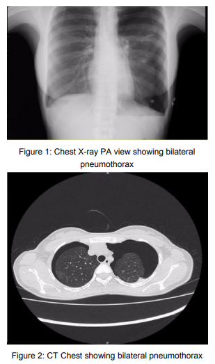
Category

volume2-issue2

427 views

Editorial

By | volume2-issue2 | No Comments

1,135 views

Journal of the Association of Pulmonologist of Tamil Nadu. Vol. 2, Issue 1, May – August 2019

EDITORIAL

Asthma Phenotypes

Asthma is a heterogenous disease. Its manifestations are different at different times to different or same stimuli. It is well known that no known asthmatics behave in the same manner to a given stimulus. Two decades ago asthma was recognised as an allergic or atopic disease. This appears to be a too simplistic explanation as global asthma patterns are not the same everywhere.

Until recently asthma was considered as an atopic disease starting in childhood, a IgE mediated Th2 inflammation characterised by eosinophilic inflammation that responds to corticosteroids in inhalants or other forms depending on the severity. Eosinophilic inflammation can be demonstrated in nasal smears, blood eosinophilia and eosinophils in endobronchial biopsies. There could be an elevation of biological markers like IgE, periostin etc., This assumption is being increasingly challenged and disproved by demonstration of noneosinophilic inflammation and alternate inflammatory pathways. Patients may have severe asthma or persistent asthma but without demonstrable eosinophils in sputum, nasal smears and bronchial wash. Repeated assesements of airways have demonstrated reproducible non eosinophilic inflammation in both short term and long term but the evidence is equivocal.

As of now asthma phenotypes are of clinical and inflammatory types. The clinical asthma phenotypes are allergic, late onset, Aspirin induced respiratory disease, obesity and GERD related asthma. The inflammatory asthma types are allergic asthma, eosinophilic asthma and non eosinophilic types of asthma. The splitting of clinical and inflammatory phenotypes is going to further increase as our knowledge about pathophysiology of asthma keeps growing.

The purpose of this phenotyping is to personalise the treatment options and individualise the treatment. The one fits all may not be good for asthma too. A high IgE type may respond to Omalizumab and high eosinophil type may respond to Benralizumab. A non eosinophilic cough variant asthmatic may respond better with LAMA (Long acting muscarinic antagonist). The placing of invasive interventions like bronchial thermoplasty also may arise. In summary it is good time for asthma patients as the basket is growing and our knowledge in pathophysiology is also growing. Probably the day is not far off when every asthmatic would have a tailor made personal prescription for better control.

In this issue an attempt has been made to phenotype the clinical types in a tertiary care hospitals with reliance on bronchial wash and the presence of Galactaman assay in Broncho alveolar lavage, blood eosinophils, serum galactaman assay. I only hope this will kindle interest in this field and come out with data.

Prof. Dr. Narasimhan R, MD FRCP (E & G)
Editor in Chief Journal of the Association of Pulmonologist of Tamil Nadu

How to cite this article : Narasimhan.R, Editorial, JAPT 2019: 2(2):42

Access this article online

Quick Response Code: Website:
www.aptchest.com
529 views

Etiology Underlying Pleural Effusion in Thanjavur Medical College Hospital: A Descriptive Study

By | volume2-issue2 | No Comments

694 views

K Anbananthan et al.: Etiology Underlying Pleural Effusion in Thanjavur Medical College Hospital: A Descriptive Study

Etiology Underlying Pleural Effusion in Thanjavur Medical College Hospital: A Descriptive Study

K Anbananthan1 , A Manimaran2 , G Karthick3, P Vanithamani3

1Associate Professor, 2 Senior Resident, 3 Junior Resident, Department of Thoracic medicine, Thanjavur Medical College and Hospital, Thanjavur, Tamil Nadu, India.

Abstract

Background: Pleural effusion is one of the most common signs seen in respiratory pathologies. An attempt to establish common etiologies underlying pleural effusion helps in effective management of the same.
Materials and Methods: After obtaining proper informed consent, patients presenting with pleural effusion underwent clinical examination in addition to radiological and biochemical investigations. Where needed, the diagnosis was confirmed using pleural biopsy and bacteriological analysis.
Results: Investigations confirmed tuberculosis in 23 patients, malignancy in seven cases, congestive cardiac failure in four cases, parapneumonic causes in 12 patients, hypoproteinemia in two patients, and pulmonary thromboembolism in two patients.
Angiogram: shows multiple feeders from right internal mammary artery supplying the sequestrated segment
of lung.
Treatment: Embolisation of two major feeding branches from right internal mammary artery done. Patient
became asymptomatic.

Corresponding Author: Dr. R. Ravikumar, Department of Radiology, Apollo Main Hospitals

How to cite this article: K Anbananthan, A Manimaran, G Karthick, P Vanithamani, Etiology Underlying Pleural Effusion in Thanjavur Medical College Hospital: A Descriptive Study, JAPT 2019: 2(2):44-46

Introduction

A pleural effusion represents the disruption of the normal mechanisms of formation and drainage of fluid from the pleural space. Pleural effusions are associated with diseases of varied etiologies and often carry a grave prognosis.1 Thus, a pleural effusion is abnormal excessive collection of fluid in pleural cavity resulting from excess fluid formation or decreased absorption2,3

Pleural effusion is classified as exudative and transudative on the basis of Light’s criteria. According to these criteria, all exudates have at least one of the following while transudates have none.

  • Ratio of pleural fluid protein to serum protein >0.5.
  • Ratio of pleural fluid lactate dehydrogenase (LDH) to serum LDH >0.6.
  • Pleural fluid LDH > 2/3 of the upper limit of serum LDH.4.

Worldwide, exudative effusions are usually due to empyema, malignancy, tuberculosis, pulmonary embolism, and connective tissue diseases.4,5 In our setup, the common causes of exudative pleural effusions are tuberculosis, parapneumonic effusion, and malignancy6,7.

rapneumonic effusion, and malignancy6,7 The relative frequency of the cause of pleural effusion is known to vary in different parts of world8. However, in developing nations, infections – especially tuberculosis and parapneumonic effusions, are more prevalent9.

Objectives

The aim of this study was to find out the etiological basis of pleural effusion in patients presenting with pleural effusion in Thanjavur Medical College Hospital.

Materials and Methods

This descriptive study was conducted in the Department of Thoracic Medicine, Thanjavur Medical College Hospital, Thanjavur, over a period of 12 months from January 2018 to December 2018. Following proper clinical examination, the underlying cause of pleural effusion was established using pleural biopsy, radiological, biochemical, cytological, and bacteriological methods. Where necessary, one or combination of many investigations was used to confirm diagnosis. About 50 patients presenting with pleural effusion were involved in the study. Patients were informed about the study and proper informed consent was given by them.

Observation and Results

It was observed that of the 50 patients presenting with pleural effusion, investigations confirmed tuberculosis in 23 patients, malignancy in seven cases, congestive cardiac failure in four cases, parapneumonic causes in 12 patients, hypoproteinemia in two patients, and pulmonary embolism in two patients.

Confirmed Diagnosis Based on Combination of Investigations As seen in Table 1, based on combination of investigations, 23 cases were confirmed as tuberculous pleuritis, 7 cases were malignancies, 12 cases were parapneumonic, 4 cases were Congestive cardiac failure, 2 cases were due to hypoproteinemia and a further 2 cases were confirmed as pulmonary embolism. This is clearly illustrated in Figure 1.

Sidedness of Pleural Effusion

As seen in Table 2, 25 cases had right sided pleural effusion, 19 cases had left sided effusion, while only 6 cases had bilateral effusion. This is clearly illustrated in Figure 2.

Discussion

This prospective study was carried out to establish the most common causes for pleural effusion.

Of 50 patients, 31 (62%) were male, whereas 19 (38%) were female with an approximate malefemale ratio of 3:2, In our study, tuberculosis was the leading cause of pleural effusion accounting for 46% of cases. This is in concordance with many such studies conducted in developing countries such as Iraq, Ghana, and Pakistan[9-11].

Most of the patients in the present study had right-sided pleural effusion (50%) which is fairly comparable with the study of Ambethiya (right side pleural effusion – 60%) and Dambal et al. (right side pleural effusion – 58.2%).[12,13] Tuberculous pleural effusion more commonly occurs in the right side because it involves the right lung more than the left lung. Majority of pleural effusions were right sided then followed by left sided and bilateral pleural effusion. These results are comparable to a study done in Ethiopia[14].

In our study, parapneumonic effusion and malignancy respectively come next in frequency as the causes. This is similar to a study done in Lahore.[6,7] Parapneumonic effusion occurred in 24% of patients which is higher compared to results from an international study by Zablockis and Nargela[4] which showed parapneumonic causes being responsible for only 13% of effusions. Malignancy accounted for 14% of our cases which is similar to the studies done by Ambethiya (malignancy – 18%) and Dambal et al.[12,13].

Pleural effusions in patients with congestive heart failure are typically bilateral. In our study, CCF accounted for only 8% of cases. Hypoproteinemia and pulmonary thromboembolism are less frequent with each accounting for 4% of cases in our study.

Conclusion

Tuberculosis is the leading cause of pleural effusion in our study. This is similar to what is  being seen in many studies conducted across developing countries. Hence, we conclude that intensive antitubercular measures may go a long way in bring down the number of patients presenting with pleural effusion.

References

1. Chowdhury PK, Ahmed S, Alam SM, Ghosh DK, Biswas SP. Etiological basis of pleural effusion in a teaching hospital. Bang Med J Khulna 2016;49:27-305.
2. Diaz-Guzman E, Dweik RA. Diagnosis and management of pleural effusions: A practical approach. Compr Ther 2007;33:237-46.
3. Frew AJ, Holgate ST. Respiratory Disease. Kumar and Clark Clinical Medicine. 6th ed. London: W.B. Saunders; 2005. p. 952-3.
4. Zablockis R, Nargela R. Diagnostic value of pleural fluid cytologic examination. Medicina (Kaunas) 2002;38:1171-8.
5. Anwar R, Faros JI. Causes of lymphocytic exudative pleural effusion as revealed by percutaneous pleural biopsy: Experience from Peshawar. Pak J Med Sci 2005;21:39-43.
6. Shaikh SJ, Memon SA. The etiology of pleural effusion in children: Hyderabad experience. Pak J Med Sci 2007;23:86- 7.
7. Anwar R, Farooqi JI. Incidence of malignancy in case of lymphocytic exudative pleural effusion, as revealed by percutaneous pleural biopsy. Med Channel 2005;11:59-61.
8. Storey DD, Dines DE, Coles DT. Pleural effusion. A diagnostic dilemma. JAMA 1976;236:2183-6.
9. Afful B, Murphy S, Antunes G, Dudzevicius V. The characteristics and causes of pleural effusions in Kumasi Ghana a prospective study. Trop Doct 2008;38:219-20.
10. Al-Alusi F. Pleural effusion in Iraq: A prospective study of 100 cases. Thorax 1986;41:492-3.
11. Khan Y, Zia SB. Etiological spectrum of exudative Pleural effusion in a tertiary care hospital based on closed pleural biopsy. Ann Pak Inst Med Sci 2011;7:133-6.
12. Kataria YP, Khurshid I. Adenosine deaminase in the diagnosis of tuberculous pleural effusion. Chest 2001;120:334-6.
13. Dambal A, Patil BS, Hegde AC. A Dissertation Submitted to Karnataka University; 1998.
14. Desalew M, Amanuel A, Addis A, Zewdu H, Jemal A. Pleural effusion: Presentation, cause and treatment outcome in a resource limited area, Ethiopia. Health 2012;4:15-19.

536 views

Phenotyping of Adult Asthmatics Visiting Pulmonology OPD of A Tertiary Care Hospital in South India

By | volume2-issue2 | No Comments

694 views

Amal Johnson et al.: Phenotyping of Adult Asthmatics Visiting Pulmonology OPD of A Tertiary Care Hospital in South India

Phenotyping of Adult Asthmatics Visiting Pulmonology OPD of A Tertiary Care Hospital in South India

Amal Johnson1, Sumi M Sam2, Narasimhan R3

1Post graduate – Department of Respiratory Medicine, Apollo Hospitals, Greams Road, Chennai, India
2Physician Assistant – Department of Respiratory Medicine, Apollo Hospitals, Greams Road, Chennai, India
3Senior Consultant Pulmonologist, Apollo Hospitals, Greams Road, Chennai, India

Abstract

Background: Asthma is a chronic inflammatory airway disease with different presentation, disease
progression, and response to therapy. However, despite this recognition, the treatment approaches for
asthma have been uniformly applied irrespective of these differences. Methods: This study is a cluster analysis done in 100 consecutive patients with Asthma diagnosed according to recent GINA guidelines visiting Pulmonology OPD in Apollo Hospitals,Chennai from January 2019 to March 2019. Results: Most patients were having Atopic asthma(75%) with history of atopy, family history of asthma and elevated Total IgE. The predominant inflammatory cell type was eosinophils(35%) followed by paucigranulocytic. Breathing difficulty and wheeze were prominent symptoms. There were 16 asthmatics who smoked currently. Six patients
diagnosed as Sensitizer induced Occupational asthma (4 cement exposure, 1 coal exposure, 1 chalk dust
exposure) and 1 patient diagnosed RADS. One patient had exacerbation prone asthma and one patient had
corticosteroid dependent/resistant asthma. Conclusion: This study is an elementary attempt to categorize
diagnosed adult asthmatics into various clusters to aid in better understanding of pathophysiological process
of asthma and to tailor specific management plan for each subtype.

Keywords: Asthma, disease, phenotype, onset, atopic

Corresponding Author: Dr. Amal Johnson, Post graduate, Department of Respiratory Medicine, Apollo Hospitals, Greams Road, Chennai, India

How to cite this article: Amal Johnson, Sumi M Sam, Narasimhan R, Phenotyping of Adult Asthmatics Visiting Pulmonology OPD of A Tertiary Care Hospital in South India, JAPT 2020: 2(2):47-54

Introduction

Asthma is a heterogeneous disease characterized by respiratory symptoms such as wheeze, breathing difficulty, chest tightness and cough that vary over time, in intensity, together with variable expiratory airflow limitation1. The
chronic respiratory disease affects 1-18% of the population2-4. The episodic flare in asthma is triggered by various stimuli, such as allergens, climate change, respiratory infections, exercise and irritants. The pathophysiology of asthma is complex involving airway inflammation, intermittent airflow obstruction and bronchial hyperresponsiveness1.
Phenotype is defined as “The composite, observable characteristics of a disease, resulting from interaction between its genetic make-up and environmental influences, which are relatively stable, but not invariable, with time”. The purpose of phenotyping of asthma is to primarily understand the pathophysiology and to cluster patients that will respond to a specific treatment5.

Review of Literature

A retrospective study conducted by JL Kuhlen et al6 entitled “Identification of Asthma Phenotypes
in a Tertiary Care Medical Center” pubished in Americal Journal of Medical Sciences (2014) in 139 consecutive patients from January 2010 to May 2012 clustered patients with asthma into 5 groups based on SARP(Severe Asthma Research Program Simple Algorithm) by Moore et al7. as following 1) Pediatric onset type, atopic type light asthma 2) Pediatric onset, atopic asthma 3) Adult-onset type, primarily nonatopic type 4) Pediatric onset type, atopic type, serious asthma 5) Adult-onset type, atopic type, serious asthma.
In study entitled “Cluster analysis and clinical asthma phenotypes” published by Halder et al8 in
Americal Journal of Respiratory and critical care medicine in 2008, he categorized asthma into five phenotypes as following 1) Early onset atopic asthma 2) Obese, noneosinophilic asthma 3) Benign asthma 4) Early symptom predominant asthma 5) Inflammation-predominant asthma.

Objectives

To categorize diagnosed adult asthmatics visiting OPD of Pulmonology in a tertiary care hospital into various multiple phenotypes based on age of onset, difference in triggers, pattern of airflow obstruction, response to treatment, frequency of exacerbation, co morbidities and type of inflammation.

Materials and Methods

STUDY TYPE: Cluster analysis
SAMPLE SIZE: 100 consecutive patients with Asthma diagnosed according to GINA guidelines (2019) visiting OPD of Pulmonology
PLACE OF STUDY: Apollo Hospitals, Greams Road, Chennai
DURATION OF STUDY: January 2019 to March 2019
EXCLUSION CRITERIA: Patients were excluded if Age 12% and 200ml). Demographic and clinical data collected included Age, Gender, BMI, age of asthma onset, personal history of allergies(allergic rhinitis/atopic dermatitis), family history of asthma/allergies, smoking history, history of asthma triggers including exercise and NSAIDS use, occupation history, history of exacerbations, history of
respiratory infections, history of comorbidites, drug history including frequency of rescue inhaler use and oral corticosteroids with dose and duration, lung function by PFT, FENO, Serum neutrophil, Absolute eosinophil count and Total IgE.

The various phenotypes for categorization used in the study are
1. Atopic asthma
2. Late onset asthma including asthma in the elderly
3. Aspirin induced Asthma
4. Infection induced Asthma
5. Smoking asthmatic excluding AsthmaCOPD overlap.
6. Exercise induced Asthma
7. Exacerbation prone Asthma
8. Persistent airflow obstruction including reversible restrictive airway disease
9. Obese asthmatic excluding Asthma-COPD overlap
10. Work related asthma – occupational asthma, irritant induced asthma
11. Symptom based phenotype – SOB predominant, wheeze predominant, cough predominant
12. Corticosteroid dependent and resistant asthma
13. Inflammatory phenotype – Eosinophilic, Neutrophilic, Mixed granulocytic, paucigranulocytic

Results

Demographics
Age – Ranged from 18 to 88 with mean of 40 years
Sex – 54 males and 46 females
BMI – Ranged from 16.6 to 41.1 with average of 26.1 kg/mm<sup>2</sup>. BMI was >30 in 13 patients.
Atopic and Non Atopic Asthma
Out of the 100 patients, 75 patients were Atopic asthma and 25 were Non Atopic asthma.
In patients with Atopic asthma, childhood history of asthma in 27 patients (36%), history of atopy (Allergic rhinitis/Atopic dermatitis) in 70(93%) patients and family history of Asthma present in 36(48%) patients. There was a male predominance (60%). Total IgE was elevated in 40(53%) patients with Atopic asthma. FeNO >25 in
all patients and FeNO >50 in 5 patients. In non Atopic Asthma, there was a female predominance(70%) with FeNO > 25 in 50% of patients. Total IgE was elevated in 2 patients.
Inflammatory phenotypes
In our study, based on the serum neutrophil and absolute eosinophil count patients were classified into inflammatory cell predominant phenotype as follows

Eosinophilic – 35%
Neutrophilic – 28%
Mixed cellular – 2%
Paucigranulocytic – 35%

Sputum based phenotype
SOB predominant – 46%
Wheeze predominant – 46%
Cough predominant – 8%

Other relevant phenotypes

Other relevant phenotypes
There were 16 asthmatics who smoked currently. Six patients diagnosed as Sensitizer induced Occupational asthma (4 cement exposure, 1 coal exposure, 1 chalk dust exposure) and 1 patient diagnosed RADS. One patient developed
asthma post tuberculosis therapy. No exercise induced asthma and aspirin induced asthma cases were noted in the study group.
One patient had exacerbation prone asthma and one patient had corticosteroid dependent/resistant asthma.

Discussion

Asthma is a chronic inflammatory disease of the airways manifested as variable airflow obstruction with symptoms like cough, shortness of breath and chest tightness.
Characterization of asthma phenotypes is important in order to
1. better understand the etiological mechanisms of asthma
2. identify specific causes
3. guide the development of new therapeutic measures that will be effective for all asthmatic patients
4. enable better management and prevention of asthma9

Phenotypes

  • Atopic asthma
  •  Late onset asthma including asthma in the elderly
  •  Aspirin induced Asthma
  •  Infection induced Asthma
  • Smoking asthmatic excluding AsthmaCOPD overlap.
  •  Exercise induced Asthma
  •  Exacerbation prone Asthma
  •  Persistent airflow obstruction including reversible restrictive airway disease
  •  Obese asthmatic excluding Asthma-COPD overlap
  •  Work related asthma – occupational asthma, irritant induced asthma
  •  Symptom based phenotype – SOB predominant, wheeze predominant, cough predominant
  •  Corticosteroid dependent and resistant asthma
  •  Inflammatory phenotype – Eosinophilic, Neutrophilic, Mixed granulocytic, paucigranulocytic

Atopic Asthma and Non atopic asthma10

This is the most common phenotype of asthma and the onset is typically during childhood/adolescence with male predominance. It is a Th2 dominant inflammatory process with both airway and blood eosinophilia. This phenotype of
asthma usually accompanies history of allergic rhinitis/dermatitis with family history of allergy. This subtype responds well to steroids and omalizumab.
In contrast to atopic asthma, there is a late onset, female predominance without allergic sensitization. Most of the patients do not give personal or family history of allergy. This phenotype is more severe than allergic asthma and less responsive to steroids.
In our study, Out of the 100 patients included, 75 patients were Atopic asthma and 25 were Non Atopic asthma.
In patients with Atopic asthma, childhood history of asthma in 27 patients (36%), history of atopy (Allergic rhinitis/Atopic dermatitis) in 70(93%) patients and family history of Asthma present in 36(48%) patients. There was a male predominance (60%). Total IgE was elevated in 40(53%) patients with Atopic asthma. FeNO >25 in
all patients and FeNO >50 in 5 patients.
In non Atopic Asthma, there was a female predominance (70%) with FeNO > 25 in 50% of patients. Total IgE was elevated in 2 patients.
This is in concordance with the results of Moore et al7. where Atopic allergic asthma was seen in 82% of patients and almost all patients had elevated IgE. FeNO was >35 in Atopic asthma whereas it is around 10-12 in non atopic asthma. There was a female premodinance in severe asthmatics both in atopic and non atopic type.

Aspirin Induced Asthma(AIA)12

This phenotype is associated with HLA-DW2 and DPB1. The usual age of onset of symptoms is 20-35 years. Patients with ASA experience rhinorrhea and nasal congestion followed by severe bronchospasm within 2 hours after ingestion of NSAID aspirin. The prevalence of AIA is 21 % according to study by Varghese et al.30 but we did not have any patients with AIA.

Infection induced asthma13

In this phenotype, the respiratory infection influences asthma in association with onset of disease/exacerbation of disease/persistence of disease. In our study, one patient developed asthma post tuberculosis therapy.

Smoking asthmatics14, 15

Patients with asthma who smoke have severe symptoms, accelerated decline in lung function, quality of life and impaired therapeutic response to corticosteroids and theophylline. Smoking greatly modifies inflammation that is associated with asthma by various mechanisms. The prevalence of smoking in asthmatics is around 25% according to
Parasuramalu et al31 and was 16% in our study.

*Criteria for diagnosis of Asthma-COPD overlap

Major –

  • Age >=40 years
  • Post bronchodilator FEV1/FVC =10 pack years of tobacco or equivalent indoor or outdoor air pollution
  • Documentation of asthma before age 40 years or BDR of >=400ml in FEV1

Minor –

  • Documentation of allergic rhinitis or atopic disease
  • BDR >=200ml and 12% from baseline values on two or more encounters
  • Peripheral serum eosinophil counts >=300 cells/ml

If all major and at least one minor criteria are present, it is termed positive.

Exercise induced asthma(EIA)16

Due to evaporation of heat and water loss from the airway Starts within 3-5mins with peak bronchoconstriction occurring at 10-15 minutes.
Usually defined as >=10% decrease in FEV1 after exercise. Exercise induced asthma may be followed by refractory perior of 4 hours during which repeated exercise causes less bronchoconstriction. Symptoms may be relieved
with beta agonist therapy. The prevalence of EIA was 5-20% according to Weiler et al32. but we did not see any patients with EIA in our study.

Exacerbation prone asthma17-19

This phenotype is characterized with history of severe exacerbation requiring an emergency room visit or hospitalization atleast once in 3 months. The increased exacerbation is independent of asthma control. On stable condition, they were mild-moderate asthmatics rather than severe asthmatics.
We had one exacerbation prone asthma in our study compared to Global prevalence of 10% according to study by Stanojevic et al33.

Persistent airflow obstruction20

This is a unique phenotype characterized by reduced FEV1/FVC in a patient receiving treatment with high dose ICS during stable phase of disease for at least 4 weeks. The decreased response to short acting bronchilators is due to airway remodeling caused by chronic inflammation of airways.
We had one patient under this subtype in our study compared to the global prevalence of 7.5% according to Roche et al34.

*Risk factors for persistent airflow obstruction –

Earlier age of onset
Longer disease duration
More severe asthma
Non atopic disease/absence of rhinitis
Very frequent exacerbations
Smoking
More inflammatory cells in sputum
Greater smooth muscle hypertrophy

Importance of earlier identification of this subtype is to avoid misdiagnosis of COPD and intensive follow-up of the patients for prompt management.

Inflammatory phenotypes21

Based on the predominant inflammatory cell involved in bronchitis, asthma can be phenotyped into aucigranulocytic, eosinophilic, neutrophilic and mixed granulocytic. Eosinophilic asthma (40%) is early onset allergic asthma (Th2 mediated inflammation) and responsive to steroids and biological agents . Non eosinophic asthma is severe
asthma phenotype (Th17 mediated inflammation) characterized by poor symptom control and corticosteroid resistance.
In our study, based on the serum neutrophil and absolute eosinophil count patients were classified into inflammatory cell predominantphenotype as follows
Eosinophilic – 35%
Neutrophilic – 28%
Mixed cellular – 2%
Paucigranulocytic – 35%
In our study, 35% patients were eosinophilic and paucigranulocytic each with neutrophilic(28%) followed by mixed cellular. This is in concordance with SARP analysis by Moore et al7.

Obese Asthma22-24

Obese asthma is characterized by early onset allergic asthma complicated by development of obesity in later age. The asthma is worsened by intra abdominal and chest wall mass which causes increased pressure on the diaphragm and chest wall causing lower tidal volume hindering bronchodilator action.
According to GC forte et al35, the prevalence of BMI >30 in asthmatics is around 13% and this is in concordance with our study.

Work related asthma25-27

  • Occupational asthma

1. Sensitizer induced asthma
2. RADS(Reactive airway dysfunction
syndrome)

  • Work exacerbated asthma

*Occupational asthma
Sensitizer induced asthma – appears after a latency period of sensitization to the causal agent
RADS (Reactive airway dysfunction syndrome) – Clinical criteria

  • Documented absence of preceding respiratory complaints
  • Onset of symptoms occurred after a single specific exposure to irritant
  • High concentration of irritant exposure(gas/fume/spray)
  • Onset 3 months
  • Symptoms simulated asthma with cough, wheeze and dyspnea predominant
  •  Spirometry show reversible airflow obstruction
  • Methacholine challenge test was positive
  • Other types of pulmonary diseases were ruled out

*Work exacerbated asthma – Patients with pre existing asthma developed worsened symptoms when exposed to occupational allergens.
In our study, six patients diagnosed as Sensitizer induced Occupational asthma (4 cement exposure, 1 coal exposure, 1 chalk dust exposure) and 1 patient diagnosed RADS. This is in concordance with results of RD Caldaeira et al where prevalence of work related asthma is 4.2%.

Symptom based phenotype28

SOB phenotype – respond well to ICS
Cough predominant – respond well to Montelukast
Wheeze predominant – respond well to ICS + Montelukast

In our study, breathing difficulty and wheeze were predominant symptoms followed by cough predominant in only 8% of patients as in study by Zedan et al28.

Corticosteroid dependent and corticosteroid resistant asthma29

Corticosteroid resistant asthma is defined as <=15% improvement in FEV1 after 2 weeks of oral prednisolone therapy 40 mg once daily. Corticosteroid dependent asthma is defined as 15-30% improvement in FEV1 after 2 weeks of oral
prednisolone therapy 40 mg once daily. Patients experience cushingoid side effects due to high dose steroid use and experience withdrawal symptoms along with worsening of symptoms and lung function after stepping down or temporary stoppage of steroid therapy.
In our study we had one patient with corticosteroid dependent asthma compared to 5-10% by SK Lukhadia et al29.

Conclusion

Asthma is not a single disease but rather a syndrome with various presentations and differential response to same treatment. There is no current guideline evidence for treatment based on phenotype so far. Future direction for research in this aspect will assist in making individualized specific treatment plan and better control of the
disease. This study is an elementary attempt to categorize diagnosed adult asthmatics into various clusters based on onset, difference in triggers, pattern of airflow obstruction, response to treatment, frequency of exacerbation, co-morbidities and type of inflammation.

Acknowledgment

We sincerely thank the management of Apollo Hospitals for the constant support in our pursuit of excellence.

References

1. 2020 GINA Main Report – Global Initiative for Asthma – GINA. https://ginasthma.org/gina-reports/
2. Masoli M, Fabian D, Holt S, Beasley R. The global burden of asthma: executive summary of the GINA Dissemination
Committee report. Allergy 2004;59:469-78.
3. Van Wonderen KE, Van Der Mark LB, Mohrs J, Bindels PJE, Van Aalderen WMC, Ter Riet G. Different definitions in childhood asthma: how dependable is the dependent variable? Eur Respir J 2010;36:48-56.
4. Godar M, Blanchetot C, de Haard H, Lambrecht B, Brusselle G. Personalized medicine with biologics for severe type 2 asthma: current status and future prospects. MAbs. 2017;10(1):34-45.doi:10.1080/19420862.2017.1392425
5. Lai CKW, Beasley R, Crane J, Foliaki S, Shah J, Weiland S, International Study of A, et al. Global variation in the prevalence and severity of asthma symptoms: phase three of the International Study of Asthma and Allergies in
Childhood (ISAAC). Thorax 2009;64:476-83
6. Kuhlen JL et al. Identification of Asthma Phenotypes in a Tertiary Care Medical Center. Am J Med Sci.2014;348(6):480-485.
7. Moore et al. National Heart, Lung, and Blood Institute’s Severe Asthma Research Program. Am J Respir Crit Care
Med. 2010 Feb 15; 181(4):315-23.
8. Halder PH, Pavord ID, Shaw DE et al. Cluster analysis and clinical asthma phenotypes. Am J Resp Crit Care Med.
2008;178:218-24.
9. Pembrey L, Barreto ML et al. Understanding asthma phenotypes: the World Asthma Phenotypes(WASP). ERJ Open Res 2018; 4: 00013-2018.
10. Saglani S, Bush A. Asthma, Atopy, and Airway Inflammation. Am J Respir Crit Care Med. 2008;178(5):437-
438. doi:10.1164/rccm.200805-796ed
11. Wüthrich B (ed): The Atopy Syndrome in the Third Millennium.Curr Probl Dermatol. Basel, Karger, 1999, vol
28, pp 129-134
12. Hamad AM, Sutcliffe AM, Knox AJ. Aspirin-induced asthma: clinical aspects, pathogenesis and management.
Drugs. 2004;64(21):2417-32.
13. Darveaux J, Lemanske RF et al. Infection related asthma. J Allergy Clin Immunol Pract. 2014 Nov-Dec; 2(6): 658–663.
14. Thomson NC, Chaudhuri R et al. Asthma and cigarette smoking. Eur Respir J 2004;24: 822-833.
15. Chatkin J, Dullius C. The management of asthmatic smokers. Asthma Res Pract. 2016;2(1). doi:10.1186/s40733-
016-0025-7
16. SD Giacco, D Firinu et al. Exercise and asthma: an overview. Eur Clin Respir J 2015;2:10.3402
17. NJ Sathish et al. Repeated exacerbation of asthma: An intrinsic phenotype of uncontrolled asthma. Lung India
2019 March;36(2):131-138
18. Corren J. Exacerbation-prone asthma-intrinsic to severe disease or a unique phenotype?. Clinical & Experimental Allergy. 2014;44(2):152-153. doi:10.1111/cea.12256
19. RJ Loymans et al. Exacerbation-prone Asthma: A seperate bioclinical Phenotype. American Journal of Respiratory and Critical Care Medicine, 195(3), pp. 275–277
20. Persistent Airway Obstruction in Asthma. Am J Respir Crit Care Med. 2013;187(1):1-2. doi:10.1164/rccm.201211-1966ed
21. Sarath S, P Nair. Asthma Endotypes and an overview of targeted therapy for Asthma. Frontiers in Medicine
2017;4:158
22. Baffi C, Winnica D, Holguin F. Asthma and obesity: mechanisms and clinical implications. Asthma Res Pract.
2015;1(1). doi:10.1186/s40733-015-0001-7
23. Dixon A, Poynter M. Mechanisms of Asthma in Obesity. Pleiotropic Aspects of Obesity Produce Distinct Asthma
Phenotypes. Am J Respir Cell Mol Biol. 2016;54(5):601-608.doi:10.1165/rcmb.2016-0017ps
24. Baffi C, Winnica D, Holguin F. Asthma and obesity: mechanisms and clinical implications. Asthma Res Pract.
2015;1(1). doi:10.1186/s40733-015-0001-7
25. Vandenplas O, Malo J. Definitions and types of workrelated asthma: a nosological approach. European
Respiratory Journal. 2003;21(4):706-712. doi:10.1183/09031936.03.00113303
26. Dykewicz et al. Occupational asthma: Current concepts in pathogenesis, diagnosis and management. Journal of
allergy and clinical immunology.March 2009;123(3):519-528
27. M Brooks S. Irritant-Induced Asthma and Reactive Airways Dysfunction Syndrome (RADS). Journal of Allergy
& Therapy. 2014;05(03). doi:10.4172/2155-6121.1000174
28. Zedan M, Attia G, Zedan M et al. Clinical Asthma Phenotypes and Therapeutic Responses. ISRN Pediatr.
2013;2013:1-7. doi:10.1155/2013/824781
29. SK Ludadia. Steroid Resistant Asthma. Journal of assofication of physicians of India. 2014; 62:38-40.
30. Varghese, M., & Lockey, R. F. (2008). Aspirin-Exacerbated Asthma. Allergy, Asthma & Clinical Immunology, 4(2), 75.
31. Parasuramalu B G, Huliraj N, Rudraprasad B M, Prashanth Kumar S P, Gangaboraiah, Ramesh Masthi N R. Prevalence
of bronchial asthma and its association with smoking habits among adult population in rural area. Indian J Public Health 2010;54:165-8
32. Weiler JM, et al. American Academy of Allergy, Asthma & Immunology Work Group report: exercise-induced asthma. J. Allergy Clin. Immunol. 2007;119:1349–1358. doi:10.1016/j.jaci.2007.02.041.
33. Stanojevic S, Moores G, Gershon AS, Bateman ED, Cruz AA, et al. Global asthma prevalence in adults: Findings
from the cross-sectional world health survey. BMC Public Health. 2012;12:204.
34. Roche, N., Dalmay, F., Perez, T., Kuntz, C., Vergnenegre, A., Neukirch, F., Huchon, G. (2008). Impact of chronic airflow obstruction in a working population. European Respiratory Journal, 31(6), 1227–1233.
35. GC Forte et al. Prevalence of obesity in asthma and its relations with asthma severity and control. rev assoc med bras. 2013;59(6):594–59

381 views

Fascioliasis – Pleuropulmonary Manifestations an Overview

By | volume2-issue2 | No Comments

694 views

Dakshinamurthy B et al.: Fascioliasis – Pleuropulmonary Manifestations an Overview

Fascioliasis – Pleuropulmonary Manifestations an Overview

Dakshinamurthy B1, Ajay Narasimhan2, Narasimhan R3

1Post Graduate, 3Senior Consultant, Dept of Respiratory Medicine, Apollo Hospitals, Chennai
2Consultant, Dept of Cardiothoracic Surgery, Apollo Hospitals, Chennai

Abstract

Fascioliasis, which is a zoonotic infestation caused by the trematode Fasciolia hepatica (liver fluke), is primarily a disease of herbivorous animals such as sheep and cattle. Humans become accidental hosts through ingesting uncooked aquatic plants such as watercress.
It presents a wide spectrum of clinical pictures ranging from fever, eosinophilia and vague gastrointestinal symptoms in the acute phase to cholangitis, cholecystitis, biliary obstruction, extrahepatic infestation, or asymptomatic eosinophilia in the chronic phase. Because of its infrequent and protean presentation and the lack of clinical data, the management of acute infection is challenging. Lung manifestations of fasciolia are very rarely reported in the literature. This article reviews the pleuropulmonary complications of fasciolia hepatica infection in humans.

Corresponding Author: Dr.Dakshinamurthy B, Department of Respiratory Medicine, Apollo Main Hospitals, 21, Greams Lane, Off Greams Road, Chennai, Tamil Nadu, India.

How to cite this article: Dakshinamurthy B, Ajay Narasimhan , Narasimhan R, Fascioliasis – Pleuropulmonary Manifestation an Overview, JAPT 2019: 2(2):55-60

Introduction

Fascioliasis is a cosmopolitan parasitic zoonosis produced by the trematode Fasciolia hepatica in its adult stage that affects herbivorous mammals (cattle, goats, and sheep) and occasionally human beings as incidental hosts1.

Fasciolia hepatica is a non-segmented flat hermaphrodite trematode, measuring 2-3 cm long by 10-13 mm wide2. Human beings are infected by consuming aquatic vegetables contaminated with metacercarias (larval form encysted and resting), particularly wild watercress (nasturdium officinale), besides mint, alfalfa, reeds, lettuce and spinach. Other sources of infection are: ingesting badly cooked liver of infected animals and, on a lesser scale, drinking contaminated water.

The biological cycle can be in one of two forms:
A. Sexual: which occurs in the definitive host (ruminants, other animals and humans).
B. Asexual: which takes place in the intermediary host (molluscs of the genera fossarea and lymnaea)3, 4.

Larvae come out of the cyst in the duodenum to penetrate through the intestinal wall and reach the peritoneum. Larvae proceed to the liver capsule and parenchyma and ultimately to the bile ducts5,6.

Parasites may rarely locate ectopically in the lungs, skin, heart, brain, stomach, eyes andlymph nodes through hematologic, lymphatic route or direct invasion7-12. On the other hand, even without a parasite localized in chest cavity, pulmonary abnormalities such as infiltrates and pleural effusions (atypical lung involvement) may occur indirectly through parasitic infection, especially in acute fascioliasis.

When we reviewed the literature, we did not encounter publications, except a few case reports, regarding lung involvement of FH13-16. Indeed, because FH is localized in the liver, signs and symptoms associated with the disease of this organ are forefront. Therefore, the findings associated with lung and other organs may be ignored by clinicians. Therefore, lung abnormalities associated with FH are not investigated with chest radiography in routine clinical practice. In our institute we came across an interesting scenario wherein the patient is a 35 year old male from Assam, a farmer by occupation presented with complaints of fever, cough with minimal expectoration and breathlessness for 3 months duration. He also had an associated right sided chest pain for the past 3 months and loss of weight of around 5 kgs in the same period. He denied prior history of anti-tuberculous treatment and was a non-smoker and an occasional alcohol user. He was evaluated elsewhere for the same complaints and found to have a right sided pleural effusion and came to our centre for further management. His chest X-ray revealed a right sided moderate pleural effusion and CT chest was also done that showed large right sided pleural effusion with fissural extension and pleural thickening, with patchy right lower lobe consolidation , calcified enlarged mediastinal lymphnodes and an evolving abscess in the superior aspect of segment 8 of liver.

Pleural fluid aspiration was done which on cytology showed sheets of eosinophils, was negative for malignant cells and negative for AFB smear and GeneXpert for MTB complex. His blood investigations were normal except for increased ESR and peripheral eosinophilia of 35% eosinophils in the differential counts. Fibre optic Bronchoscopy was done that was normal study and negative for GeneXpert for MTB complex.

Fig 1 – Thorascopic Image Showing Visceral and Parietal
Pleural Nodules and Adhesions

Fig 2 : Right Pleural Effusion with ICD in SITU

Hence we proceeded with rigid thoracoscopy which revealed multisepated pleural effusion with lots of adhesions and multiple nodules in the parietal and visceral pleura [FIG 1] Intercostal drainage tube was placed in situ [FIG 2]. Biopsy of the pleura showed fibroblastic proliferation with intense eosinophilia suggestive of eosinophilic pleuritis. Hematologist opinion was also sought wherein which a bone marrow aspiration cytology showed trilineage haematopoiesis, normoblastic maturation, eosinophilia and plasmacytosis and increased iron stores. Bone marrow trephine biopsy showed mildly hyper cellular marrow with eosinophilia, mild plasmacytosis and increased iron stores. Moving ahead we proceeded with Contrast CT of the abdomen that showed branching linear tracts over the surface of liver into the substance of liver on both the lobes very much suggestive of FASCIOLIA HEPATICA [FIG 3] infection involving the right pleura, liver and also the lower pole of the right kidney. The patient was started on T.Nitazoxanide 500 mg thrice daily for 2 weeks and the Intercostal tube was removed after the lung expanded adequately and drain was minimal.

FIG 3 : Fasciolia Tunnel Sign – Linear Tracts Over the Liver Surface

Going retrospectively we enquired his eating habits and found that he used to take fresh raw uncooked vegetable salad grown from his kitchen backyard and also had the habit of drinking liquor with fresh water fish dipped in it and reciprocally the scenario was like we had to “fish out the fasciolia from the pleural wine”.

Around 7% of the pleural effusion are eosinophilic in nature with more than 10% of eosinophils in the differential counts 17,18. Mostly the pleural fluid eosinophilia is due to either air or blood in the pleural space. The factor responsible for the increased colony-forming activity of eosinophils and their increased survival appears to be IL-5 19,20, although IL-3 and granulocyte or macrophage colony-stimulating factor (GM-CSF) may also play a role (19). The source of the IL-5 appears to be the CD4 lymphocyte in the pleural fluid (20), but the eosinophils in the pleural fluid may themselves also produce IL-5 (19). The source of the eosinophils in eosinophilic pleural effusions appears to be the bone marrow; no progenitor cells are present in the pleural space. The most common cause of pleural fluid eosinophilia is air in the pleural space. In a series of 127 cases with more than 20% eosinophils in the pleural fluid, 81 (64%) were thought to have pleural fluid eosinophilia secondary to air in the pleural space (21).The second most common cause of pleural fluid eosinophilia is blood in the pleural space. Following traumatic hemothorax, pleural fluid eosinophils do not usually become numerous until the second week (21). There is frequently an associated peripheral blood eosinophilia that does not disappear until the pleural effusion is completely resolved (22). The pleural effusions associated with pulmonary embolization are frequently bloody and contain numerous eosinophils (23). Bloody pleural fluids due to malignant disease are not usually characterized by eosinophilia (21) Kalomenidis and Light (24) reviewed the etiology of 392 cases of eosinophilic pleural effusions when cases associated with pleural air and/or blood were excluded. They reported that the most common diagnosis was idiopathic (39.8%), followed by malignancy (17%), parapneumonic (12.5%), transudates (7.9%), tuberculosis (5.6%), pulmonary embolism (4.3%), and others (12.8%). In a recent study of 13 5 patients with eosinophilic pleural effusions from a single institution, the following diseases were responsible: malignancy 34.8%, infections 19.2%, unknown 14.1%, posttraumatic 8.9%, and miscellaneous 23.0% (17). Pleural eosinophilia is common in patients with asbestos-related pleural effusions. Patients with eosinophilic pneumonia frequently have pleural effusions and the mean eosinophil percentage in the pleural fluid was 38% in one study (25). The pleural effusions secondary to drug reactions are frequently eosinophilic. Offending drugs include dantrolene, bromocriptine, and nitro-furantoin. Pleural effusions secondary to parasitic diseases such as paragonimiasis (26), hydatid disease (27), amebiasis (21), or ascariasis (21) frequently contain a large percentage of eosinophils. Lastly, the pleural effusion associated with the Churg-Strauss syndrome is also eosinophilic (28).

Acute fascioliasis refers to the initial, hepatic phase of the parasitic disease. It is characterized by tissue destruction caused by the migration of the immature parasites from the small intestine to the biliary system. Because of its invasive nature, the acute stage of fascioliasis causes far more morbidity than chronic infection when adult worms inhabit the biliary tree. However, as for other neglected diseases, comprehensive clinical studies are lacking, and the most convincing clinical data derive from small retrospective case series or single case reports [29-32]. The typical clinical spectrum of acute fascioliasis encompasses anorexia (sometimes associated with nausea and vomiting), which might lead to weight loss, abdominal pain, especially in the upper abdomen, tender hepatomegalia, night sweats, fever, and other immunemediated manifestations, such as urticaria or arthralgias. Other, less frequently reported, findings include splenomegalia, ascites, subcapsular hepatic haematoma, intra-abdominal haemorrhage, pleural or pericardial effusion, and respiratory symptoms [33,31,34]. Ectopic larvae in other localizations, such as subcutaneous tissue, eyes, and the central nervous system,may cause a variety of other clinical manifestations31. The leading laboratory abnormality is hypereosinophilia; hypergammaglobulinaemia and anaemia might also be present35. The incubation period ranges from a few days to several weeks; the acute stage of fascioliasis usually lasts for 2–4 months33.

Patients with respiratory symptoms in the foreground are evaluated as atypical fascioliasis (31). Cough, hemoptysis and chest pain may be seen in these patients (36). Some patients have eosinophilrich sputum discharge and dyspnea and they are erroneously treated as asthma (37, 38). In various studies, complaint of cough was found as 14%, 15% and 33% in the acute phase (31, 39, 40). Arigona et al. reported chest pain and dyspnea in 3 out of 20 fascioliasis patients (31). In another study, respiratory symptoms were detected in 10 patients (17.9%) and three patients with lung involvement belonged to this group (41). They detected cough in 14.2% patients, dyspnea in 12.5%, chest pain in 8.9% and sputum in 3.6%. Co-existence of cough, dyspnea and chest pain in two out of three patients is striking. Simultaneous chest radiographs were not obtained in other studies, thus lung involvement or presence of primary pulmonary disease that may have been a component of fascioliasis could not be evaluated. Pulmonary signs and symptoms are considered to be related with hepatic destruction caused by larvae and inflammatory response. The severity of symptoms has been reported to vary according to number of parasites, location, immune response of host against parasite and especially eosinophil level(31) . Larvae were reported to ectopically locate in the lungs, skin, heart, brain, stomach, epididymis, eyes and lymph nodes(7-12). Ectopic fascioliasis appears as masses and abscesses with eosinophilic and mononuclear infiltrations develop due to tissue injury in these organs(21). How parasites migrate to ectopic tissues is not known. However, different theories suggest that migration may occur through hematogenous, lymphatic or direct route(22,23). Presence of larvae in the thorax may only be detected with thoracoscopy and biopsies and these procedures are not recommended for definite diagnosis due to their invasive nature. Physical examination findings of the lungs vary according to the pathology caused by FH.

Usually the rales are presence when cough is in the foreground. Dullness on percussion, is detected when a pleural effusion develops and a tympanic sound is detected in the presence of a pneumothorax. A decrease is detected in respiratory sounds on auscultation. Bronchial sounds and rales may be heard when infiltration and consolidation develop. Lung involvement may be seen in different forms in radiologic evaluation. Loeffler syndrome like displacing parenchymal infiltrates and pleural effusion are the most common radiological findings (13). However, publications are available reporting pneumothorax development (16). Routine laboratory test results of the patients who developed lung abnormalities (white blood count, ESR, ALT, AST, ALP and bilirubin levels) were similar to those of the patients without lung abnormality. An eosinophilic reaction may be shown in the fluid obtained in presence of pleural effusion and pericarditis (31).

Hepatic imaging is crucial in patients with possible acute fascioliasis. In concordance with other published data29,30,31, abdominal ultrasound did not contribute to the diagnosis of acute fascioliasis. However, this technique is of value in chronic fascioliasis, when adult worms inhabit the biliary tract, causing inflammation and/or dilatation, and might be seen as mobile structures in the gall bladder or choledochus45. A useful imaging technique is CT, which reveals hypodense focal lesions that might be confused with metastasis or abscess; more typical for acute fascioliasis are hypodense tunnel-like lesions, (fasciolia tunnel sign ) which, over time, slowly change in a centripetal manner32. Magnetic resonance imaging, which offers no diagnostic advantage, is characterized by hypointense lesions in T1- weighted images, and hyperintense signals in T2 images35. Periportal lymph nodes are not infrequent but are non-specific. The combination of upper abdominal pain, marked eosinophilia and hypodense lesion in CT imaging is highly indicative of acute fascioliasis35. Other rare causes of this combination of findings include hepatic toxocariasis and hepatic capillariasis.

The easiest means of establishing the diagnosis is visualization/demonstration of ova in the stool (2), but it has low sensitivity. Because of no ova release in the stool in the acute phase and intermittent ova release in the chronic phase of the disease, serological tests may be necessary to confirm the diagnosis.

Triclabendazole is the current treatment of choice for fascioliasis but unfortunately the drug is not available in india. It is administered at a dose of 10 mg/kg/day either once or twice over two consecutive days. The other alternative drug is T nitazoxanide 500 mg twice daily for 2 weeks.

Conclusion

The Fasciolia hepatica transmission chain is well documented with 70% of infections related to the consumption of watercress by persons living in the high risk areas. Consequently, when undertaking the clinical evaluation of patients from these high risk areas with abdominal pain and eosinophilia, one should consider the possibility of a Fasciolia hepatica infection and, for the clinical evaluation, include questions of the patient’s food consumption habits as well as performing the relevant faeces tests and Fasciolia hepatica tests. Fascioliasis should be kept in mind in the differential diagnosis and treatment of patients with respiratory symptoms and eosinophilia. Once the presence of Fasciolia hepatica infection has been established and successfully treated, then it is also important to ensure long term follow-up checks to control for any possible long term effects, possibly irreversible, that could have arisen from the Fasciolia hepatica infection

References

1. Atias A, Neghme A (1991) Fascioliasis in Parasitología Clínica. Publication’s Tecnicas Mediterráneo, Santiago, Chile, pp. 334-340.
2. Berenguer J Mosby/Doyma Libros. (2nd Edn), Elsevier SA, Netherlands. pp. 800-802.

3. Alban M, Jave J, Quisipe T (2002) Fascioliasis en Cajamarca. Revista de Gastroenterología del Perú 22 pp. 28-32.

4. López M, Huerto L, Hernandéz S, Acuña A, Nari A (1996) Fasciolosis en la Republica Oriental de Uruguay 12: 37-43. 5.

5. King CH. Flukes (liver, lung and intestinal). In: Kliegman RM, Behrman RE, Jenson HB, StantonBF (eds). Nelson Textbook of Pediatrics (18th ed). Philadelphia: WB Saunders Elsevier, 2007: 1510-1511.

6. Aksoy DY, Kerimoğlu U, Oto A and et al. Infection with fasciolia hepatica. Clin Microbiol Infect 2005; 11: 859-61.

7. Zali MR, Ghaziani T , Shahraz S and et al. Liver, spleen, pancreas and kidney involvement by human fascioliasis: imaging findings. BMC Gastroenterology 2004; 4: 15.

8. Basarali MK, Kaplan I, Cicek M and Cakir F. The relationship between trace elements and ceruloplasmin with severity of fascioliasis patients. ActaMedicaMediterranea, 2013, 29: 177- 81.

9. Pulpeiro JR, Armesto V, Varela J and et al: Fascioliasis: findings in 15 patients. Br J Radiol 1991; 64: 798-801. 10. Öngören AU, Ozkan AT, Demirel AH and et al. Ectopic intraabdominal fascioliasis. Turk J Med Sci 2009; 39: 819-23.

11. Xuan LT, Thien Hun N, Waikagul J. Cutaneous fascioliasis: a case report in Vietnam. Am J Trop Med Hyg 2005; 72: 508-9.

12. Cheng AC, Zakhidov BO, Babadjonova LJ and et al. A 6- year old boy with facial swelling and monocular blindness. Clin Infect Dis 2007; 45: 1238-9.

13. Aliaga L, Díaz M, Quiroga J and et al. Eosinophilic pulmonary disease caused by Fasciolia hepatica. Description of a case and review of the literature. Med Clin (Barc) 1984; 82: 764- 7.

14. Corredoira JC, Pérez R, Casariego E and et al. Eosinophilic pleural effusion caused by Fasciolia hepatica. EnfermInfeccMicrobiolClin 1990; 8: 258-9.

15. Ibáñez de Maeztu JC, Aguirrebengoa K, Montejo M and et al. Pleural effusion associated with acute hepatic fascioliasis. EnfermInfeccMicrobiolClin 1994; 12: 320-1.

16. Aghajanzadeh M, Sarshad A, Ebrahimian R. Pneumothorax a rarity in fascioliasis. w w w . a m s . a c . i r / A I M /9924/aghajanzadeh9924.html

17. Krenke R, Nasilowski J, Korczynski P, et al. Incidence and etiology of eosinophilic pleural effusion. Eur Respir J 2009; 34:1111-1117.

18. Ferreiro L, San Jose E, Gonzalez-Barcala FJ, et al. Eosinophilic pleural effusion: incidence, etiology and prognostic significance. Arch Bronconeumol. 2011;47;504- 509.

19. Nakamura Y, Ozaki T, Kamei T, et al. Factors chat stimulate the proliferation and survival of eosinophils in eosinophilic pleural effusion: relationship co granulocytemacrophage colony-stimulating factor, interleukin-5, and interleukin-3. Am J Respir Cell Mo! Biol. 1993;8:605-611

20. Schandene L, Namias B, Crusiaux A, et al. IL-5 in posttraumatic eosinophilic pleural effusion. Clin Exp Immunol. 1993;93:115-119.

21. Spriggs AI, Boddingcon MM. The Cytology of Effesions, 2nd ed. New York, NY: Grune& Scraccon;l968

22. Maltais F, Laberge F, Cormier Y. Blood hypereosinophilia in the course of post-traumatic pleural effusion. Chest. 1990; 98:348-351.

23. Romero Candeira S, Hernandez Blasco L, Soler MJ, et al. Biochemical and cycologic characteristics of pleural effusions secondary co pulmonary embolism. Chest. 2002;121:465-469.

24. Kalomenidis I, Light RW. Eosinophilic pleural effusions. Curr Opin Pulm Med. 2003;9:254-260.

25. Philit F, Etienne-Mastroianni B, Parrot A, et al. Idiopathic acute eosinophilic pneumonia: a study of 22 patients. Am J Respir Crit Care Med. 2002;166:1235-1239.

26. Klein A, Talvani A, Cara DC, et al. Stem cell factor plays a major role in the recruitment of eosinophils in allergic pleurisy in mice via the production of leukotriene B 4′ J Immunol. 2000; 164:4271-4276

27. Yacoubian HD. Thoracic problems associated with hydatid cyst of the dome of the liver. Surgery. 1976;79:544-548

28. Erzurum SE, Underwood GA, Hamilos DL, et al. Pleural effusion in Churg-Strauss syndrome. Chest. 1989;95: 1357- 1359.

29. Graham CS, Brodie SB, Weller PF. Imported Fasciolia hepatica infection in the United States and treatment with triclabendazole. Clin Infect Dis 2001; 33: 1–5.

30. Noyer CM, Coyle CM, Werner C, Dupouy-Camet J, Tanowitz HB, Wittner M. Hypereosinophilia and liver mass in an immigrant. Am J Trop Med Hyg 2002; 66: 774– 776.

31. Arjona R, Riancho JA, Aguado JM, Salesa R, Gonza´lezMarcı´as J. Fascioliasis in developed countries: a review of classic and aberrant forms of the disease. Medicine (Baltimore) 1995; 74: 13–23.

32. Marcos LA, Tagle M, Terashima A et al. Natural history, clinicoradiologic correlates, and response to triclabendazole in acute massive fascioliasis. Am J Trop Med Hyg 2008; 78: 222–227.

33. World Health Organization. Report of the WHO Informal Meeting on use of triclabendazole in fascioliasis control. Geneva: WHO, 2007.

34. MacLean JD, Cross J, Mahanty S. Liver, lung and intestinal fluke infections. In: Guerrant RL, Walker DH, Weller PF, eds. Tropical infectious diseases: principles, pathogens & practise, 2nd edn. Philadelphia, PA: Churchill Livingston Elsevier, 2005; 1349–1369.

35. Marcos LA, Terashima A, Gotuzzo E. Update on hepatobiliary flukes: fascioliasis, opisthorchiasis and clonorchiasis. Curr Opin Infect Dis 2008; 21: 523–530.

36. Schussele A, Laperrouza C. Les distomatoses hepatiques: a propos de 9 observations person) Facey RV, Marsden PD: Fascioliasis in man: Anoutbreak in Hampshire. Br Med J 1960; 2: 619- 25

37. Tropical and Geographical Medicine. 2nd ed. New York: Mc Graw-Hill, 1990: 473-89.

38. Flores M, Merino J, Aguirre C: Pulmonary infiltrates as first sign of infection by Fasciolia hepatica.Eur J Respir Dis 1982; 63: 231-3.

39. Karahocagil.MK, Akdeniz H, Sünnetçioğlu M and et al. A familial outbreak of fascioliasis inEastern Anatolia: A report with review of literature. ActaTropica 2011; 118: 177-83.

40. Turhan O, Korkmaz M, Saba R and et al. Seroepidemiology of fascioliasis in the Antalyaregion and uselessness of eosinophil count as asurrogate marker and portable ultrasonographyfor epidemiological surveillance. Infez. Med 2006; 14: 208-12.

41. Cengizhan sezgi Pulmonary Findings In Patients With Fascioliasis .Acta Medica Mediterranea, 2013, 29: 841

42. Catchpole BN, Snow D. Human ectopic fascioliasis. Lancet 1952; 2: 711-12

43. Rushton B, Murray M. Intrahepatic vascular lesions in experimental and natural ovine fascioliasis.J Pathol 1978; 125: 11-6.

44. Neff GW, Dinavahi RV, Chase V and et al. Laparoscopic appearance of fasciolia hepaticainfection. GastrointestEndosc 2001; 53: 668-71.

45. Cosme A, Ojeda E, Cilla G et al. Fascioliasis hepatobiliar. Estudio de una serie de 37 pacientes. Gastroenterol Hepatol 2001; 24: 375–380.

409 views

Ubiquitous Fungus Irking Inclined Wheezers – A succinct on Allergic Bronchopulmonary Aspergillosis

By | volume2-issue2 | No Comments

694 views

Amal Johnson et al.: Ubiquitous Fungus Irking Inclined Wheezers – A succinct on Allergic Bronchopulmonary Aspergillosis

Ubiquitous Fungus Irking Inclined Wheezers – A succinct on Allergic Bronchopulmonary Aspergillosis

Amal Johnson1, Vaseema Thabassum2, Narasimhan R3

1Post graduate –Department of Respiratory Medicine, Apollo Hospitals, Greams Road, Chennai, India
2Post graduate –Department of Respiratory Medicine, Apollo Hospitals, Greams Road, Chennai, India
3Senior Consultant Pulmonologist, Apollo Hospitals, Greams Road, Chennai, India

Abstract

Aspergillus mold cause respiratory disease depending upon the host immunity and organism virulence. ABPA is the most significant manifestation of allergic aspergillosis. It is an immunological pulmonary disease caused by hypersensitivity reaction to the aspergillus antigen in susceptible patients, most commonly with asthma and cystic fibrosis. The presentation can range from mild symptoms to extensive lung disease that may manifest as respiratory failure. This review summarizes the history, epidemiology, risk factors, immunopathogenesis, pathology, clinical features, diagnostic criteria, laboratory and radiological investigations and management of ABPA.

Keywords: ABPA, Asthma, Aspergillus, IgE

Corresponding Author: Dr.Amal Johnson, Post graduate, Department of Respiratory Medicine, Apollo Hospitals, Greams Road, Chennai,
India

How to cite this article: Amal Johnson, Vaseema Thabassum, Narasimhan R, “Ubiquitous Fungus Irking Inclined Wheezers” A succinct on Allergic Bronchopulmonary Aspergillosis, JAPT 2019: 2(2):61-67

Introduction

Aspergillus mold cause respiratory disease depending upon the host immunity and organism virulence. The manifestations can be classified into 3 distinct clinical categories; allergic aspergillosis (Allergic Aspergillus sinusitis, ABPA), saprophytic colonization (Aspergilloma) and invasive aspergillosis (Airway invasive aspergillosis, chronic necrotizing aspergillosis and invasive aspergillosis)1. ABPA, the most significant manifestation of allergic aspergillosis is immunological pulmonary disease caused by hypersensitivity reaction (Type 1,3, 4) to the aspergillus fumigatus antigen(Af) in susceptible patients mostly commonly with asthma and cystic fibrosis2,3(Cf).

History of ABPA

ABPA was first described in 1952 by Hinson, Moon and Plummer described three patients with recurrent wheezing, pulmonary infiltrates, eosinophilia in blood and sputum, and brown plugs or flecks in expectorated mucus4.

Epidemiology of ABPA

The prevalence of ABPA in asthma varied between 2.5% and 22.3% according to the International Society for Human and Animal Mycology (ISHAM) working group. Most cases present in the 3rd to 5th decade5.

Genetic Factors Involved in ABPA

Genetic risk factors include expression of HLA-DR2 and HLA-DR5 genotypes, while HLADQ2 protected against ABPA6,7. In subjects with CF, increased chances of Aspergillus colonization of the airways and subsequent development of ABPA were found in those with CF transmembrane conductance regulator gene mutations8,9. Surfactant protein-A2 polymorphisms, elevated levels of mannan-binding lectin due to the 1011A allele, and toll-like receptor polymorphisms 43, IL -4,IL-10,1L13,1L-15 polymorphisms also play an important role in the development of ABPA10,11,12.

Pathology of ABPA

Histologic examination reveals the presence of mucus, fibrin, Curschmann spirals, CharcotLeyden crystals, and inflammatory cells with scanty hyphae. The bronchial wall in ABPA is usually infiltrated by eosinophils and other inflammatory cells. Bronchocentric granulomatosis, the presence of noncaseating granulomas containing eosinophils and multinucleated giant cells centered on the airway is also a feature14, 15, 16.

Pathogenesis of ABPA

Clinical Features of ABPA

The presentation can range from mild asthma, with very few symptoms, to extensive lung disease that may manifest as respiratory failure. Patients encounter frequent exacerbations and if left untreated, lead to chronic fibrotic lung disease. Respiratory symptoms included cough (99%), breathlessness (99%), expectoration (98%), wheezing (97%), and haemoptysis (41%). Nasal symptoms suggestive of upper airways allergy were present in 45%. Expectoration of sputum plugs was reported by 37% of the patients and nasal plugs by 6%. Approximately half of the patients had a personal/family history of atopy.

Physical examination in ABPA may reveal rhonchi and crepitations. These patients may also exhibit cyanosis, digital clubbing, and features of corpulmonale in end stage fibrotic stages(15%)17.

Diagnostic Criteria

Staging of ABPA:

Investigations

1) Serum Eosinophil Count

Absolute eosinophil count greater than 1000 cells/ul is one of the major criteria for ABPA diagnosis. However, low eosinophil counts does not rule out ABPA24.
2) Skin Testing with Aspergillus Antigens

Aspergillus skin testing is done using commercial or locally prepared Aspergillus fumigatus antigen. The test is read every 15 minutes for 1 hour and then after 6-8 hours. The reactions are classified as Type 1: If wheal and erythema develops within 1 min, reaches maximum within 10-20 minutes and resolves within 1-2 hours and Type 3: Is read after 6 hours and if edema present, termed as positive17.

3) Sputum Cultures for A fumigatus

Culture of A. fumigatus in the sputum is supportive but not diagnostic of ABPA. The fungus can also be grown in patients with other pulmonary diseases due to the ubiquitous nature of the fungi17.

4) Total Serum IgE

The total IgE level is the most useful test for diagnosis and follow-up of ABPA. A normal serum IgE level excludes ABPA as the cause of the patient’s current symptoms. After treatment with glucocorticoids, the serum IgE levels decline, and a >35 % is taken as a criteria for remission. The serum IgE determination is also used for follow-up, and a doubling of the patient’s baseline IgE levels indicates relapse of ABPA25

5) Pulmonary Function Tests

These tests help categorize the severity of the lung disease but have no diagnostic value in ABPA and need not constitute the basis for screening. The usual finding is an mixed obstructive with restrictive defect of varying severity26-29. Normalisation of the parameters can be noticed after treatment.

6) Precipitating Antibodies against A. fumigatus 

By the double gel diffusion technique, precipitating antibodies against Af could be detected in the serum upto 92% of patients in concentrated serum30,31.

7) Radiological manifestations

Differential Diagnosis13

1. Aspergillus hypersensitive bronchial asthma
2. Tuberculosis/CAP
3. Inflammatory pulmonary disorders – eosinophilic pneumonia, bronchocenteric

Complications13

1) Recurrent asthma exacerbations
2) Bronchiectasis
3) Pulmonary hypertension
4) Respiratory failure
5) End stage fibrotic lung disease

Treatment

The goals of treatment of ABPA according to the AAAAI Committee report on ABPA32 are

1) Control symptoms of asthma or CF2) Prevent or treat pulmonary exacerbations of ABPA
3) Reduce or remit pulmonary inflammation
4) Mitigate progression to end stage fibrotic
or cavitatory disease.

a) Oral Steroids

Oral corticosteroids is the treatment of choice for ABPA.

Steroid dosing in ABPA

Stage 1(Acute) and Stage 3(Exacerbation)33

Prednisolone 0.5mg/kg/day for 2 weeks followed by alternate days for 6-8 weeks. Once Ig declines by 1/3rd and radiological opacities cleared, tapered 2.5 to 5mg every 2 weeks. After completion of therapy, patient monitored for 2 months to assure remission.

Stage 4 (steroid dependent asthma) –

Alternate therapy with prednisolone 10- 40mg/day to maintain symptom control34

Stage 5 (Fibrotic lung disease) – management of end stage lung disease

Pulse therapy with intravenous methylpred nisolone using 10 to 20 mg/kg/day for 3 consecutive days has been shown to be useful in managing severe and sometimes life-threatening exacerbations among children with ABPA and cystic fibrosis35

b) Antifungals (azole)

Antifungals reduce the fungal load thereby decreasing the antigenic stimulus and inflammatory response and proven to have adjunctive role in steroid dependent or steroid unresponsive patients36. They are also used in initial therapy of acute and exacerbation of ABPA37. The mechanism of action is by blocking the CYP450 dependent demthylation of lanosterol, leading to the inhibition of ergosterol synthesis. Ergosterol depletion enchances fungal cell susceptibility to both oxidative and nonoxidative phagocytic damage. The most common used drug is itraconazole followed by newer azoles, voriconazole and posaconazole. Itraconazole has low oral bioavailability and high side effect profile. Dose adjustments are required for patients with mild to moderate hepatic dysfunction(Child class A and B). Monitoring of LFT is recommended at baseline, within the first 2 weeks of treatment initiation and periodically thereafter. The dosage of itraconazole recommended is 200 mg twice daily for 4 to 6 months, which is then tapered over the next 4 to 6 months. Oral itroconazole has side effect of nausea and diarrhea causes by the excipient hydroxypropyl B cyclodextrin which is used to increase the solubility of the drug. It is associated with a unique triad of hypertension, hypokalemia, and edema. Also causes negative inotropic effect avoiding administration in patients with a history of heart failure. By inhibiting steroid metabolism and thereby exacerbating adrenal suppression, itraconazole might lead to cushingoid features when used for very long durations.

Voriconazole is extended spectrum azole with good oral bioavailability and does not inhibit steroid metabolism. It is associated with visual disturbances and cutaneous phototoxicity in a minority of patients.

c) AntiIgE

Omalizumab, a monoclonal antibody against IgE has been tried and shown excellent results in patients with steroid dependent ABPA38-39.

Conclusion

All patients with uncontrolled asthma should be evaluated for allergic bronchopulmonary aspergillosis. Diagnosis of ABPA is based on clinical, immunological and radiological criteria and the management is primarily steroids with antifungals having an adjunct role.

References

1. Soubani AO, Chandrasekar PH. The clinical spectrum of pulmonary aspergillosis. Chest 2002; 121:1988 –1999

2. Rajan TV. The Gell-Coombs classification of hypersensitivity reactions: a re-interpretation. Trends Immunol 2003; 24:376 – 379

3. Geha RS, Sampson HA, Askenase PW, et al. Allergy and hypersensitivity. In: Janeway CA, Travers P, Walport M, et al. eds. Immunobiology. New York, NY: Garland, 2001; 517–556

4. Hinson, K., Moon, A., & Plummer, N. (1952). Bronchopulmonary Aspergillosis : A Review and a Report of Eight New Cases. Thorax, 7(4), 317-333

5. Agarwal R, Chakrabarti A, Shah A, Gupta D, Meis JF, Guleria R, et al. Allergic bronchopulmonary aspergillosis: review of literature and proposal of new diagnostic and classification criteria. Clin Exp Allergy 2013;43:850-73.

6. Chauhan B, Santiago L, Kirschmann DA, Hauptfeld V, Knutsen AP, Hutcheson PS, et al. The association of HLADR alleles and T cell activation with allergic bronchopulmonary aspergillosis. J Immunol 1997;159:4072- 6.

7. Chauhan B, Santiago L, Hutcheson PS, Schwartz HJ, Spitznagel E, Castro M, et al. Evidence for the involvement of two different MHC class II regions in susceptibility or protection in allergic bronchopulmonary aspergillosis. J Allergy Clin Immunol 2000; 106:723-9

8. Miller PW, Hamosh A, Macek M Jr, Greenberger PA, MacLean J, Walden SM, et al. Cystic fibrosis transmembrane conductance regulator (CFTR) gene mutations in allergic bronchopulmonary aspergillosis. Am J Hum Genet 1996;59:45-51.

9. Eaton TE, Weiner Miller P, Garrett JE, Cutting GR. Cystic fibrosis transmembrane conductance regulator gene mutations: do they play a role in the aetiology of allergic bronchopulmonary aspergillosis? Clin Exp Allergy 2002;32:756-61.

10. Saxena S, Madan T, Shah A, Muralidhar K, Sarma PU. Association of polymorphisms in the collagen region of SPA2 with increased levels of total IgE antibodies and eosinophilia in patients with allergic bronchopulmonary aspergillosis. J Allergy Clin Immunol 2003;111:1001-7.

11. Kaur S, Gupta VK, Shah A, Thiel S, Sarma PU, Madan T. Elevated levels of mannan-binding lectin [corrected] (MBL) and eosinophilia in patients of bronchial asthma with allergic rhinitis and allergic bronchopulmonary aspergillosis associate with a novel intronic polymorphism in MBL. Clin Exp Immunol 2006;143:414-9.

12. Carvalho A, Pasqualotto AC, Pitzurra L, Romani L, Denning DW, Rodrigues F. Polymorphisms in toll-like receptor genes and susceptibility to pulmonary aspergillosis. J Infect Dis 2008;197:618-21.

13. Agarwal, R. (2009). Allergic Bronchopulmonary Aspergillosis. Chest, 135(3), 805-826. doi: 10.1378/chest.08- 2586

14. Chan-Yeung M, Chase WH, Trapp W, et al. Allergic bron chopulmonary aspergillosis. Clinical and pathologic study of three cases. Chest 1971; 59:33–39

15. 66 Riley DJ, Mackenzie JW, Uhlman WE, et al. Allergicbronchopulmonary aspergillosis: evidence of limited tissue invasion. Am Rev Respir Dis 1975; 111:232– 23

16. Case records of the Massachusetts General Hospital.Weekly clinicopathological exercises. Case 24– 2001. A 46-year-old woman withchronic sinusitis, pulmonary nodules, and hemoptysis. N Engl J Med 2001; 345:443–449

17. Agarwal, R. (2009). Allergic Bronchopulmonary Aspergillosis. Chest, 135(3), 805-826. doi: 10.1378/chest.08- 2586

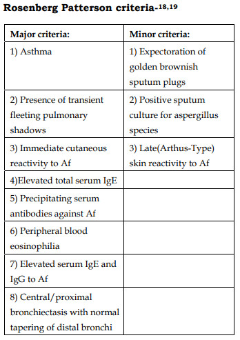
18. Rosenberg M, Patterson R, Mintzer R, et al. Clinical and immunologic criteria for the diagnosis of allergic bronchopulmonary aspergillosis. Ann Intern Med 1977; 86:405–414

19. Patterson R, Greenberger PA, Halwig JM, et al. Allergic bronchopulmonary aspergillosis: natural history and classification of early disease by serologic and roentgenographic studies. Arch Intern Med 1986; 146:916– 918

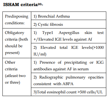
20. Agarwal R, Chakrabarti A, Shah A, Gupta D, Meis JF, Guleria R, et al. Allergic bronchopulmonary aspergillosis: review of literature and proposal of new diagnostic and classification criteria. Clin Exp Allergy 2013;43:850-73.

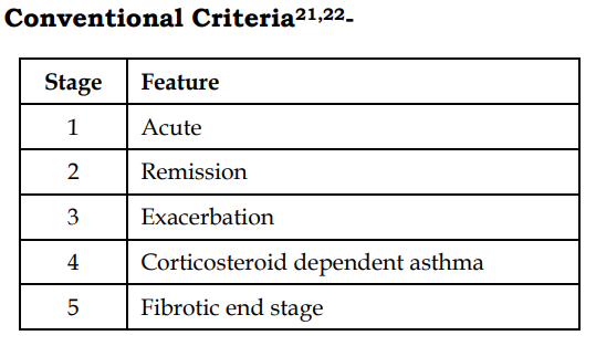
21. Patterson R, Greenberger PA, Radin RC, Roberts M. Allergic bronchopulmonary aspergillosis: staging as an aid to management. Ann Intern Med 1982;96:286-91.

22. Greenberger PA, Patterson R. Diagnosis and management of allergic bronchopulmonary aspergillosis. Ann Allergy 1986;56:444-8.

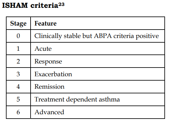
23. Agarwal R, Chakrabarti A, Shah A, Gupta D, Meis JF, Guleria R, et al. Allergic bronchopulmonary aspergillosis: review of literature and proposal of new diagnostic and classification criteria. Clin Exp Allergy 2013;43:850-73.

24. Agarwal R, Gupta D, Aggarwal AN, et al. Allergic bronchopulmonary aspergillosis: lessons from 126 patients attending a chest clinic in North India. Chest 2006; 130:442– 448

25. Ricketti AJ, Greenberger PA, Patterson R. Serum IgE as an important aid in management of allergic bronchopulmonary aspergillosis. J Allergy Clin Immunol 1984; 74:68–71

26. 115 Malo JL, Hawkins R, Pepys J. Studies in chronic allergic bronchopulmonary aspergillosis: 1. Clinical and physiological findings. Thorax 1977; 32:254–261

27. McCarthy DS, Pepys J. Allergic broncho-pulmonary aspergillosis. Clinical immunology: 1. Clinical features. Clin Allergy 1971; 1:261–286

28. Nichols D, Dopico GA, Braun S, et al. Acute and chronic pulmonary function changes in allergic bronchopulmonary aspergillosis. Am J Med 1979; 67:631–637

29. Panjabi C, Shah A. Lung functions in allergic bronchopulmonary aspergillosis. Respirology 2006;11:A38.

30. Longbottom JL, Pepys J. Pulmonary aspergillosis: diagnostic and immunological significance of antigens and C-substance in Aspergillus fumigatus. J Pathol Bacteriol1964; 88:141–151

31. Vlahakis NE, Aksamit TR. Diagnosis and treatment of allergic broncho pulmonary aspergillosis. Mayo Clin Proc 2001; 76:930–938

32. Greenberger PA, Bush RK, Demain JG, Luong A, Slavin RG, Knutsen AP. Allergic bronchopulmonary aspergillosis. J Allergy Clin Immunol Pract 2014;2:703-8.

33. Greenberger PA. Allergic bronchopulmonary aspergillosis.J Allergy Clin Immunol 2002; 110:685– 692

34. Shah, A., & Panjabi, C. (2016). Allergic Bronchopulmonary Aspergillosis: A Perplexing Clinical Entity. Allergy, Asthma & Immunology Research, 8(4), 282. doi: 10.4168/aair.2016.8.4.282

35. Thomson JM, Wesley A, Byrnes CA, Nixon GM. Pulse intravenous methylprednisolone for resistant allergic bronchopulmonary aspergillosis in cystic fibrosis. Pediatr Pulmonol 2006;41:164-70.

36. Leon EE, Craig TJ. Antifungals in the treatment of allergic bronchopulmonary aspergillosis. Ann Allergy Asthma Immunol 1999; 82:511-6.

37. Ullmann, A., Aguado, J et al. (2018). Diagnosis and management of Aspergillus diseases: executive summary of the 2017 ESCMID-ECMM-ERS guideline. Clinical Microbiology And Infection, 24, e1-e38

38. Tillie-Leblond I, Germaud P, Leroyer C, Tétu L, Girard F, Devouassoux G, et al. Allergic bronchopulmonary aspergillosis and omalizumab. Allergy 2011;66:1254-6.

39. Collins J, Devos G, Hudes G, Rosenstreich D. Allergic bronchopulmonary aspergillosis treated successfully for one year with omalizumab. J Asthma Allergy 2012;5:65-70.

370 views

Bilateral Catamenial pneumothorax with Catamenial Hemoptysis

By | volume2-issue2 | No Comments

694 views

Bilateral Catamenial pneumothorax with Catamenial Hemoptysis

Bilateral Catamenial pneumothorax with Catamenial Hemoptysis

Selvi C, Govindasamy C, Narasimhan.R

Department of Respiratory Medicine, Apollo Hospitals, Chennai, Tamilnadu, India

Abstract

Bilateral catamenial pneumothorax coexisted with catamenial hemoptysis is a rare entity. It is usually caused by thoracic endometriosis syndrome. We report a case 24 year healthy female with no comorbid status presenting with bilateral catamenial pneumothorax with catamenial hemoptysis. This lady presented with breathlessness, bilateral chest pain and streaky hemoptysis for four months associated with her menstrual cycle. Chest x ray and CT chest done showed bilateral pneumothorax. Serum CA 125 levels was elevated.USG pelvis done was normal. Bronchoscopy was done in view of hemoptysis showed bilateral inflamed mucosa no active bleeding. Videothoracoscopy was done showed multiple nodules over the diaphragm, biopsy taken from that nodules following biopsy, diaphragmatic meshplasty and talc pleurodesis was done. To prevent the recurrence patient was started on hormonal therapy. Patient improved clinically and radiologically and no recurrence till one year of followup.
Keywords: Breathlessness, Hemoptysis, Pneumothorax, Videothoracoscopy, Meshplasty ,Pleurodesis

Corresponding Author: Dr.Narasimhan.R, Department of Respiratory Medicine, Apollo Main Hospitals, 21, Greams Lane, Off Greams Road, Chennai – 600 006, Tamil Nadu, India.

How to cite this article: Selvi C, Govindasamy C, Narasimhan.R, Bilateral Catamenial pneumothorax with Catamenial Hemoptysis, JAPT 2019: 2(2):68-71

Introduction

Catamenial pneumothorax (CP) is a recurrent spontaneous pneumothorax occurring in women associated with a period of menstruation. Catamenial pneumothorax occurs due to the presence of ectopic endometrial tissue in thoracic cavity.1,2. Endometriosis of thoracic cavity is a rare entity; it consists of four clinical types: Catamenial pneumothorax, Catamenial hemothorax, hemoptysis and pulmonary nodules 3,4. CP usually occurs within 24 hours before and 72 hours after the onset of menses and appears almost exclusively in the right hemithorax1. Around 60% of thoracic endometriosis cases are associated with pelvic endometriosis1,5. Cause for thoracic endometriosis

is not clear but most acceptable theory explains the retrograde implantation of endometrial tissue by lymphatic and haematogenous dissemination or presence of diaphragmatic defects 2. Upto 85 to 90% of cases of CP usually occurring on right side. A combined surgical and hormonal treatment is the management of choice for CP. Hormonal therapy prevent the postoperative recurrences.3,6. We report a catamenial pneumothorax occurring bilaterally along with catamenial hemoptysis which was successfully treated with combined surgical and hormonal therapy.

Case Report

24 year old healthy woman presented with symptoms of breathlessness and chest pain and hemoptysis for past 4 months associated with her menses. First three episode resolved spontaneously. This time she had increasing breathlessness, chest pain and hemoptysis. On presentation patient was tachypneic her respiratory rate was high, other vitals stable. On respiratory examination Bilateral decreased chest movements and decreased breath sounds was heard. Her hematological and biochemical parameters were normal. HIV serology was negative. Her CA125 level was elevated(56U/ml). Chest X-ray was done showed bilateral pneumothorax (Fig. 1). Computed tomography (CT) done showed bilateral pneumothorax (Fig. 2). Ultrasonography of pelvis excluded the presence of pelvic endometriosis

Hypothesis of catamenial pneumothorax and catamenial hemoptysis was taken into account .the patient underwent a bronchoscopy showed inflamed mucosa on both side, no active bleeding.

videothoracoscopic exploration of the bilateral pleural cavity under general anesthesia was done, first in right lateral position by two ports approach pleural cavity explored, multiple purple red nodules were seen in the diaphragm. Biopsy taken from that nodules .Patient repositioned and redraped and procedure repeated on right side, Talc pleurodesis done bilaterally followed by bilateral meshplasty of diaphragm done. After control of hemostasis a ICD drain was placed in pleural space and full lung re-expansion achieved (Fig. 3). The chest tube was removed after 2 days of surgery (Fig. 4).

Final pathology confirmed endometriosisrelated catamenial pneumothorax (ER–CP) according the most updated classification by Legras et al.8. After surgery the patient was started hormonal therapy with 17 etinil testosterone 800mg daily then shifted to dienogest 2mg daily. No recurrence has been observed till 3 months post surgery

3. Discussion

We report a rare case of bilateral Catamenial pneumothorax (CP) coexisted with catamenial hemoptysis. It is a rare entity characterized by lung collapse during menstruation due to thoracic endometriosis. CP most commonly occurs in 30 -50 years of premenopausal women with history of infertility.

Thoracic endometriosis syndrome consists of two types. Pulmonary form and pleural form. Pulmonary form manifests as catamenial hemoptysis and pulmonary nodules. Pleural endometriosis manifests as catamenial pneumothorax, catamenial hemothorax, catamenial pneumomediastinum and chest pain11. Several pathogenic mechanisms have been evoked CP. The metastatic theory presume the transdiaphragmatic passage of endometrial tissue by lymphohematogenous dissemination or congenital fenestration. The anatomical theory explains for dissolution of cervical mucus plug during the menses with migration of cells through vagina, uterus, fallopian tubes to peritoneum and air reaching the chest by diaphragmatic congenital fenestrations. In the hormonal theory prostaglandin F2 mediated pulmonary vasospasm during ovulation leads to ischemic injury and alveolar rupture4,6. Congenital diaphragmatic defects are more common on the right side and also more lymphatic drainage and clockwise peritoneal circulation that sweeps endometrial implants to right side this explains CP most commonly occur on right side11,12.

The diaphragmatic defect(s) can be single or multiple, commonly occur at the central tendon, this defects may be tiny holes (1–3 mm), or larger defects (>10mm)13. High resolution computed tomography (HRCT) to detect endometrial deposits in the lung and pleura. USG of pelvis to detect pelvic endometriosis because around 50 to80% pulmonary endometriosis associated with pelvic endometriosis.14,15. Both thoracic and pelvic MRI are considered superior to CT due to the blood products in the endometrial deposits11,16.

CA125 (Cancer Antigen 125) is a tumor marker used for monitoring treatment and detecting recurrence of ovarian cancer. It also elevated in benign ovarian conditions also. Endometriosis also been associated with increased levels of CA-125, and although it is not considered a specific marker, it can assist in early diagnosis of endometriosis-related pneumothorax 13,17.

Bagan et al conclude that this CA-125 biological marker may favor indication of videothoracoscopy and hormonal therapy at an early stage in the prevention of catamenial pneumothorax recurrences, and should be evaluated prospectively in a larger-scale studx 18.

Cyclical haemoptysis at the time of menstruation is called catamenial hemoptysis. Diagnosis by detection of endometriotic plaques on bronchial washings and endometrial tissue on lung biopsy. However, these may be negative despite the presence of the disease. Computed tomography scan of the chest during the days of menstruation and in midcycle shows ‘ground-glass’ or patchyinfiltrate appearance during the menstrual days and clear lung fields in midcycle.

Management of Thoracic endometriosis can be medical or surgical. Medical treatment using 17etinil testosterone or danazol it suppresses the production of gonadotropins, endometrial hypotrophy thus leading to a condition of pseudomenopause and finally a remission of the disease.14,20. Endometriosis is a hormone dependent disease so when medical therapy stopped disease will get recur morethan 50%within 6 months of treatment.20. The surgical treatment combined with hormonal therapy is recommended modality of treatment4,21.

Surgical treatment should be done during menstruation for better visualization of pleurodiaphragmatic endometriosis. Videothoracoscopy is a very useful tool since it assesses both the diaphragmatic surface and the pleural cavity and is able to detect multiple nodules although it has been reported, No pathologic findings were reported in 8.5%cases1,3. Suturing the defect, endoscopic resection of the area, covering the diaphragmatic surface with polyglactin mesh are the common surgical procedures but which do not completely prevent the risk of CP to recur. Pleurodesis plays a major role when microscopic endometrial foci or newly implanted lesions cannot be detected2,12. In our case we found multiple nodules in posterior diaphragmatic surface but fenestrations were detected. we preferred our surgical procedure to biopsy from the lesion and diaphragmatic meshplasty and talc pleurodesis, and combine it with the hormonal treatment to achieve the best outcome.

4. Conclusion

Coexisting bilateral Catamenial pneumothorax and catamenial hemoptysis is a rare presentation. So videothoracoscopy provides magnification and better visualization of the pleural cavity and diaphragm. Among surgical options videothoracoscopic resection of detected lesions combined with pleurodesis with diaphragmatic meshplasty is the most successful one and, associated with hormonal therapy, reduces the risk of recurrence.

References

1. A.N. Visouli, K. Darwiche, A. Mpakas, et al., Catamenial pneumothorax: a rare entity? Report of 5 cases and review of the literature, J. Thorac. Dis. 4 (S1)(2012) 17–31.

2. C. Nezhat, J. Main, C. Paka, A. Nezhat, R.E. Beygui, Multidisciplinary treatment for thoracic and abdominopelvic endometriosis, JSLS 18 (2014) 1–7.

3. S. Attaran, A. Bille, W. Karenovics, L. Lang-Lazdunski, Videothoracoscopic repair of diaphragm and pleurectomy/abrasion in patients with catamenial pneumothorax. A 9-year experience, Chest 143 (2013) 1066– 1069.

4. P. Azizad-Pinto, D. Clarke, Thoracic endometriosis syndrome: case report and review of the literature, Perm. J. 18 (2014 Summer) 61–65.

5. A. Veeraswamy, M. Lewis, A. Mann, S. Kotikala, B. Hajhosseim, C. Nazhat, Extragenital endometriosis, Clin. Obstet. Gynecol. 53 (2010) 449–466.

6. D. Papafragaki, L. Concannon, Catamenial pneumothorax: a case report and review of the literature, J. Women’s Health 17 (2008) 367–372.

7. P. Ciriaco, G. Negri, L. Libretti, et al., Surgical treatment of catamenial pneumothorax: a single centre experience, Interact. Cardiovasc. Thorac. Surg. 8 (2009) 349–352.

8. A. Legras, A. Mansuet-Lupo, C. Rousset-Jablonski, A. Bobbio, P. Magdeleinat, N. Roche, J.F. Regnard, A. Gompel, D. Damotte, M. Alifano, Pneumothorax in women of childbearing age: an update classification based on clinical and pathologic findings, Chest 145 (2014) 354–360.

9. P. Athwal, K. Patel, C. Hassani, S. Bahadori, P. Nardi, A case of multisystem endometriosis, J. Radiol. Case Rep. 7 (2013) 1–6.

10. C. Rousset-Jablonski, M. Alifano, G. Plu-Bureau, et al., Catamenial pneumothorax and endometriosis-related pneumothorax: clinical features and risk factors, Hum. Reprod. 26 (2011) 2322–2329.

11. P.S. Chatra, Thoracic endometriosis: a case report, J. Radiol. Case Rep. 6 (2012) 25–30.

12. V. Baysungur, C. Tezel, E. Okur, B. Yilmaz, Recurrent pneumothorax diagnosed as catamenial after videothoracoscopic examination of the pleural cavity, Surg. Laparosc. Endosc. Percutan. Tech. 21 (2011) 81–83.

13. A.N. Visouli, K. Zarogoulidis, I. Kougioumtzi, et al., Catamenial pneumothorax, J. Thorac. Dis. 6 (S4) (2014) 448– 460.

14. S.Z.A. Badawy, P. Shrestha, Recurrent catamenial pneumothorax suggestive of pleural endometriosis, Case Rep. Obst. Gynecol. (2014) 1–2, http://dx.doi.org/10.1155/2014/756040.

15. D. Soriano, R. Schonman, I. Gat, E. Schiff, D.S. Seidman, H. Carp, A.Y. Weintraub, A. Ben-Nun, M. Goldenberg, Thoracic endometriosis syndrome is strongly associated with severe pelvic endometriosis and infertility, J. Minim. Invasive. Gynecol. 19 (2012) 742–748.

16. P. Rousset, C. Rousset-Jablonski, M. Alifano, A. MansuetLupo, J.N. Buy, M.P. Revel, Thoracic endometriosis syndrome: CT and MRI features, Clin. Radiol. 69 (2014) 323–330.

17. P. Santulli, I. Streuli, I. Melonio, L. Marcellin, M. M’Baye, A. Bititi, B. Borghese, M.C. for severity of deep endometriosis, J. Minim. Invasive. Gynecol. 22 (2015) 275– 284.

18. P. Bagan, P. Berna, J. Assouad, M. Riquet, et al., Value of cancer antigen 125 for diagnosis of pleural endometriosis in females with recurrent pneumothorax, Eur. Respir. J. 31 (2008) 140–142.

19. P.J. Pickhardt, M.E. Hanson, Incidental adnexal masses detected at low-dose unenhanced CT in asymptomatic women age 50 and older: implications for clinical management and ovarian cancer screening, Radiology 257 (2010) 144–150

312 views

A Rare Case of Dapsone Induced Eosinophilic Pneumonia without Peripheral Eosinophilia

By | volume2-issue2 | No Comments

694 views

Ramkumar P.P et al.: A Rare Case of Dapsone Induced Eosinophilic Pneumonia without Peripheral Eosinophilia

A Rare Case of Dapsone Induced Eosinophilic Pneumonia without Peripheral Eosinophilia

Ramkumar P.P1, Mahilmaran.A2

1Department of Thoracic Medicine, Villupuram Medical College, Villupuram, Tamil Nadu.
2Department of Thoracic Medicine, Madras Medical College, Chennai, Tamil Nadu.

Abstract

Eosinophilic lung diseases are a diverse group of pulmonary disorders which may
result from different etiologies, including drug treatment. Dapsone, a sulfone drug useful for treating wide
variety of infectious and inflammatory dermatological conditions including leprosy, has been described as a
possible cause of eosinophilic lung diseases. We report a 40 year old female patient with leprosy who
presented with respiratory symptoms and pulmonary infiltrates and was diagnosed as suffering from
eosinophilic pneumonia which was managed successfully.
Keywords: Eosinophilic pneumonia, Dapsone, Leprosy.

Corresponding Author: Dr. P.P.Ramkumar, Department of Thoracic Medicine, Villupuram Medical College, Villupuram, Tamil Nadu.

How to cite this article: Ramkumar .P.P, Mahilmaran .A, A Rare Case of Dapsone Induced Eosinophilic Pneumonia without Peripheral Eosinophilia, JAPT 2019: 2(2):72-75

Introduction

Pulmonary eosinophilia can be caused by numerous drugs and may be life threatening1,2. Dapsone, a sulfone antibiotic is still used as a first line drug for Hansen’s disease. The lung is one of the organ involved in Dapsone Hypersensitivity Syndrome2. However, the frequency of lung involvement is unknown and only 14 cases have been previously reported in the literature3,4. We report a case of Dapsone Hypersensitivity syndrome with eosinophilic pneumonia without peripheral eosinophilia in Hansen’s disease.

Case Report

Case History and Examination

A 40 year old female diagnosed as lepromatous leprosy was admitted 8 weeks after initiating WHO multidrug therapy (Dapsone, Clofazamine, Rifampicin). She presented with a history of high grade fever, dyspnea, skin rashes all over the body with itching for 1 week. On admission her RR – 28 / min, PR – 110 /min, BP – 90/50 mm of Hg, body temperature -102°F and oxygen saturation was 85% in room air. She was icteric with generalized erythematous maculopapular rash predominantly involved on the face, trunk and extremities. Lung auscultation revealed bilateral diffuse crackles and wheeze

Investigations

A complete blood count revealed leucocytosis and absolute eosinophil count was 250. Her liver function test were deranged (Total bilirubin – 6.3 mg/dl, Direct bilirubin – 3.2 mg/dl , SGOT – 70 U/L, SGPT – 80 U/L, ALP – 90 U/L). ABG showed Type 1 respiratory failure (pH = 7.46, PaO2 =50.4 mmHg, PaCO2 =37.4 mmHg and bicarbonate =24.2 mmol/L under condition of room air oxygen inhalation). CT chest showed diffuse ground glass opacities with centrilobular nodules and septal thickening (Figure 1). Patient underwent bronchoscopy with bronchioalveolar lavage and transbronchial lung biopsy. Bronchioalveolar lavage fluid contained 25% eosinophils. Smear and culture of the BAL fluid was negative for infectious pathogens, CBNAAT – mycobacterium tuberculosis not detected. Transbronchial lung biopsy showed respiratory epithelial mucosa with connective tissue stroma showing abundant eosinohils and lymphocytes without evidence of granuloma. Stool examination was negative for parasites. Rheumatological evaluation were negative (ANA – negative). Echo cardiogram was normal.

Management

In view of suspected Dapsone Hypersensitivity Syndrome, multidrug therapy (Dapsone, Clofazamine, Rifampicin) was discontinued and patient was treated with oxygen therapy and high dose methyprednisolone for 3 days followed by prednisolone maintainance. Patient improved symptomatically and her liver parameters normalised after one week of treatment. Rechallenge with Rifampicin and clofazamine did not produce adverse effect. On addition of small dose of dapsone (25mg) patient developed respiratory symptoms and allergic reactions. So dapsone was stopped. Erythema Nodosum Leprosum was ruled out by dermatologist and patient was started on alternate regimen (Rifampicin, Ofloxacin, Clofazamine). The steroid was withdrawn after gradual tapering follow up CT chest (Figure 2) taken after 8 weeks showed significant resolution and patient was symptomatically better. The patient was symptomfree on a follow-up examination several months after the cessation of steroid treatment

Discussion

Eosinophilic pneumonias are a heterogeneous group of disorders characterized by varying degrees of pulmonary parenchymal and/or blood  eosinophilia. It may result from several different etiologies, including infectious, inflammatory, toxic and idiopathic1. A wide variety of drugs and toxic substances are important causes of pulmonary eosinophilic infiltrates. Of the medications implicated, many are commonly used antibiotics (nitrofurantoin, sulfa-containing antibiotics), cardiovascular medications (amiodarone) nonsteroidal anti-inflammatory agents, anticonvulsants (phenytoin), and antidepressants2. Dapsone commonly causes hypersensitivity syndrome but rarely causes eosinophilic pneumonia6,7 .

Patients with drug-induced eosinophilic lung disease can present as a simple eosinophic pneumonia-like syndrome or a fulminant, acute eosinophilic pneumonia-like syndrome2. Dapsone induced allergic reaction can manifest as Dapsone hypersensitivity syndrome (DHS) and DRESS syndrome (Drug rash with eosinophilia and systemic symptoms). The classic triad of DHS consists of fever, rash, and internal organ involvement (most commonly liver). Wheezing may be present, but obstructive physiology is not common on pulmonary function testing. Although radiographic findings are not specific, interstitial or alveolar infiltrates are typically evident on chest radiograph and chest computed tomographic findings include ground-glass opacity, consolidation, nodules, and irregular lines2,3.

The diagnosis of eosinophilic lung disease can bemade if any of the following findings is present: (1) pulmonary opacities with peripheral eosinophilia, (2) increased eosinophils in bronchoalveolar lavage (BAL) fluid or (3) tissue eosinophilia confirmed either by open or transbronchial lung biopsy5,8,9.

A diagnosis of drug induced eosinophilic pneumonia is based upon a careful review of drug exposures and other causes of eosinophilic lung disease must be excluded (1). A concurrent skin rash can support the diagnosis of drug-induced eosinophilic pneumonia. Discontinuation of drug usually leads to resolution of symptoms, pulmonary infiltrates, eosinophilia and normalization of lung function within a month. In severely ill patients corticosteroids may hasten recovery3,4,5.

Conclusion

Dapsone is a commonly implicated drug causing systemic hypersensitivity syndrome. However to the best of our knowledge pulmonary eosinophilia without peripheral eosinophilia has not been reported in leprosy patients. The primary treatment is the immediate discontinuation of the drug with the administration of systemic glucocorticoids. In eosinophilic lung disorder drug induced eosinophilic pneumonia should also be considered as differential diagnosis and a proper drug history must be obtained.

References

1. Cottin V. Eosinophilic Lung Diseases.Clin Chest Med. 2016 Sep;37(3):535-56.

2. KuruvillaM, Khan DA.Eosinophilic Drug Allergy.Clin Rev AllergyImmunol.2016 Apr;50(2):228-39.

3. H.K. Kim , N.R. Jeong , J.H. Park , H.J. Jang .A Case of Dapsone Induced Eosinophilic Pneumonia. American Journal of Respiratory and Critical Care Medicine 2018;197.

4. Adar T, Tayer-Shifman O, Mizrahi M, Tavdi S, Barak O, Shalit M. Dapsone induced eosinophilic pneumonia. Eur Ann Allergy ClinImmunol44: 144-146, 2012.

5. Kaur J, Khandpur S, Seith A, Khanna N. Dapsone-induced eosinophilic pneumonitis in a leprosy patient. Indian J Lepr77: 267-271, 2005.

6. YuheiKinehara 1, Takashi Kijima 1, Koji Inoue. Dapsone Hypersensitivity Syndrome-related Lung Injurywithout Eosinophilia in the Bronchoalveolar Lavage Fluid. Intern Med 54: 827-831, 2015

7. Arunthathi S, Raju S. Dapsone induced pulmonary eosinophiliawithout cutaneous allergic manifestations: an unusual encounter -a case report. ActaLeprol11: 3-5, 1998

8. Jaffuel D, Lebel B, Hillaire-Buys D, et al. Eosinophilic pneumonia induced by dapsone. BMJ317: 181, 1998

9. Janier M, Guillevin L, Badillet G. Pulmonary eosinophilia associated with dapsone. Lancet343: 860-861, 1994

570 views

Early Onset Chyliform Pleural Effusion-A Rare Case

By | volume2-issue2 | No Comments

694 views

Sridhar R et al.: Early Onset Chyliform Pleural Effusion-A Rare Case

Case Report

Early Onset Chyliform Pleural Effusion-A Rare Case

Sridhar R1, Rajeswari P2, Mahilmaran A2

1Department of Respiratory Medicine – Apollo Hospitals, Chennai.
2Department of Thoracic medicine – Madras Medical College, Chennai.

Abstract

Chyliform pleural effusion is turbid or milky from high lipid content without cholesterol crystals. Chyliform
pleural effusion is a rare presentation than chylous or pseudochylous pleural effusion. The mean duration of
the effusion to turn chyliform is 5 years. A 20 year old male presented with dry cough, left side chest pain and
dyspnea on exertion and gives history of bilateral pleural effusion for which he had taken empirical
antituberculous treatment. Chest radiograph showed bilateral pleural effusion more on left side. Thoracentesis
done on left side revealed milky white coloured fluid with elevated cholesterol levels and Mycobacterium
tuberculosis was detected and was not resistant to Rifampicin in cartridge based nucleic acid amplification
test[CBNAAT] for pleural fluid. Contrast enhanced CT chest showed healed fibrotic lesion in bilateral upper
lobe with bilateral pleural effusion with thickened pleura on left side. Patient diagnosed as chyliform effusion
and treated with Intercostal drainage and antituberculous treatment. Less than 200 cases of chyliform have
been reported in the medical literature of which microbiologically confirmed tuberculous chyliform effusion is
rarely reported. Chyliform effusion rarely develops within one year and Tuberculosis presenting as Chyliform
pleural effusion is a rare phenomenon.
Keywords: chyliform effusion, CBNAAT, tuberculosis

Corresponding Author: Dr.Sridhar R, Department of Respiratory Medicine – Apollo Hospitals, Chennai.

How to cite this article: Sridhar R,Rajeswari P, Mahilmaran A, Early Onset Chyliform Pleural Effusion-A Rare Case, JAPT 2019 2(2):76-79

Introduction

Pseudochylothorax is a cholesterol-rich
pleural effusion that is commonly associated with chronic inflammatory disorders such as  tuberculosis or rheumatoid arthritis1. Pleural fluid is milky or at least turbid and it persists after centrifugation, due to a high lipid content of the pleural fluid. High levels of lipid accumulate in the pleural fluid in two conditions. First, when the thoracic duct is disrupted, chyle can enter the pleural space to produce a chylous pleural effusion.
Second, pseudochylothorax is high cholesterol content and milky pleural fluid often with cholesterol crystals seen on microscopy. Chyliform pleural effusion is turbid or milky from high cholesterol content without cholesterol crystals2. Chyliform pleural effusion is a rare presentation than chylous or pseudochylous pleural effusion.
Less than 200 cases have been reported in the medical literature of which microbiologically  confirmed tuberculous chyliform effusion is rarely reported. The etiology is tuberculosis in 54% of

Case Report

A 20 year old male engineering student, smoker presented with dry cough, left side chest pain and dyspnea on exertion and gave history of empirical antituberculous treatment one month before for clinically diagnosed tuberculous bilateral pleural effusion. Patient gave the history of straw coloured fluid on pleural aspiration seven months before in some other hospital and pleural fluid reports were not available. On examination patient
was conscious, oriented, comfortable at rest, general examination was normal, stony dullness and decreased breath sounds on left infrascapular &amp; infraaxillary area. Other system examination was normal. Chest radiograph showed bilateral pleural effusion more on left side, USG confirmed the same. Diagnostic thoracentesis revealed milky  white coloured fluid and it was sent for analysis which showed

  • Triglycerides elevated (224mg/dl)
  • Cholesterol elevated (324mg/dl) levels
  • Cholesterol crystals -negative
  • CBNAAT –Mycobacterium tuberculosis detected, Rifampicin resistant not detected.
  • Pleural fluid culture both aerobic and anaerobic—no growth
  • Pleural fluid AFB smear-negative

Serum lipid profile:

  • Cholesterol-150
  •  Triglycerides-80
  •  HDL-23
  •  Pleural fluid cholesterol to triglyceride ratio >1
  •  Pleural fluid cholesterol to serum
  • cholesterol ratio >1

Contrast enhanced CT chest showed healed fibrotic lesion in bilateral upper lobe with bilateral pleural effusion and thickened pleura on left side. Bronchoscopy was done no abnormality was found in the tracheobronchial tree, bronchial wash for CBNAAT was negative. Contrast enhanced CT abdomen was normal and Rheumatoid factor was negative.

Tube thoracostomy was done on left side which drained 500 ml of milky white fluid and antituberculous  treatment started according to RNTCP guidelines and thorcostomy tube was removed after drainage stopped and
symptomatically improved.

Discussion

Milky turbid pleural fluid can be due to chylothorax, pseudochylothorax or empyema. Milky turbid fluid does not clear on centrifugation can be either chylothorax or pseudochylothorax but in empyema, it clears due to sedimentation of cellular debris. In our case it was not clear on centrifugation so it was probably either  chylothorax or pseudochylothorax2 . Milky pleural fluid with high triglycerides level favours chylothorax and high cholesterol level favours pseudochylothorax5 , in our case both were elevated. To further differentiate between these two we applied the ratio of pleural fluid triglycerides to pleural fluid cholesterol which was <1, ratio of serum cholesterol to pleural fluid cholesterol also 2 . Radiologically, thickened pleura was also more suggestive of pseudochylothorax than chylothorax because chyle is non-irritant, chylothorax is usually not  associated with pleural thickening or loculation 1 . Some authors mentioned cholesterol crystals negative pseudochylothorax as chyliform effusion2 . In our case elevated pleural fluid cholesterol, thickened pleura and negative cholesterol crystals favoured the diagnosis of chyliform effusion.

When a patient is diagnosed as a chyliform pleural effusion, the possibility of tuberculosis should always be entertained. In our case, CBNAAT of chyliform fluid confirmed tuberculosis. The diagnosis and therapeutic decision may be difficult in tuberculous pseudochylothorax. In the review of the literature by Garcia-Zamalloa et al., Ziehl-Neelsen stain was positive in only two cases, levels of adenosine deaminase correlated with infectious activity in 40% of those tested, and M. tuberculosis was isolated in 14% (11% of pleural fluid cultures and 17% of pleural biopsy cultures) 3 . The polled sensitivity of CBNAAT in pleural fluid was 51.4% and specificity was 98.6% 9 . So far no study has been published on sensitivity of CNNAAT in chyliform fluid. This is the first case report of CBNAAT positive chyliform pleural effusion

The mean duration of the effusion to turn chyliform is 5 years. Only 9% of patients with pseudochylothorax developed in pleural effusion of less than 5 years’ duration 4 ; the most frequent cause was parasitic infection followed by rheumatoid arthritis 6, 7 , carcinoma of the lung 8 . In our case chyliform effusion developed within one year which was very rare. So far only one case of tuberculous pseudochylothorax developing in a pleural effusion of less than one years’ evolution has been reported4.

Antituberculosis treatment is recommended for patients with pseudochylothorax in which  M. tuberculosis is isolated, in culture-negative patients with a history of past tuberculosis, and in purified protein derivative–positive patients with progressive effusions 3 . Decortication should be considered in chyliform effusion if the patient is symptomatic and the underlying lung is believed to be functional. In our case the patient improved with intercostal drainage and Antituberculosis treatment.

Conclusion

Milky pleural fluid with pleural thickening should consider pseudochylothorax than chylothorax which needs to be confirmed by biochemical analysis. The ratio of pleural fluid triglycerides to pleural fluid cholesterol suggestive of pseudochylothorax even triglycerides level elevated above normal. Chyliform effusion rarely develops within one year and Tuberculosis presenting as chyliform pleural effusion is a rare phenomenon which should be kept in mind in non-resolving pleural effusion with thickened pleura especially in high prevalence country like India.

References

1. Wrightson JM, Stanton AE, Maskell NA, Davies RJO, Lee YCG. Pseudochylothorax without pleural thickening: time to reconsider pathogenesis?. Chest. 2009;136(4):1144‐1147.
2. Light RW. Chylothorax and pseudochylothorax. In: Light RW, ed. Pleural Diseases. 3rd ed. Baltimore, MD: Williams& Wilkins; 1995:284–298.
3. Garcia-Zamalloa A, Ruiz-Irastorza G, Aguayo FJ, et al.Pseudochylothorax: report of 2 cases and review of the
literature. Medicine (Baltimore). 1999;78:200–207
4. Nogueras C, Monteagudo M, Vila M, Cabezuelo A,Mariscal D, Berlanga E. Recent-onset tuberculous pleurisy
presenting as pseudochylothorax. Am J Med2002;113:166-8.
5. Vaz MA, Teixeria LR, Vargas FS, Carmo AO, AntonangeloL, Onishi R, et al. Relationship between pleural fluid and serum cholesterol levels. Chest 2001;119:204-10.
6. Carel RS, Schey G, Bruderman I. Chyliform Pleural Effusion: An Unusual Manifestation of Hepatothoracic Echinococcus Cysts. Chest. 1975 Oct 1;68(4):598-9.
7. Muresan C 1 , Muresan L 2 , Grigorescu I 1 , Dumitrascu DL 1 Lung India. Chyliform effusion without pleural thickening in a patient with rheumatoid arthritis: A case report. 2015 Nov-Dec;32(6):616-9.
8. Goldman A, Burdorf TH. Cholesterol pleural effusion: a report of three cases with a cure by decortication. Dis
Chest. 1950;18: 586–594.
9. Sehgal IS, Dhooria S, Aggarwal AN, Behera D, Agarwal R. 2016. Diagnostic performance of Xpert MTB/RIF in tuberculous pleural effusion: systematic review and meta-analysis. J Clin Microbiol 54:1133–1136.

402 views

Radiology Quiz

By | volume2-issue2 | No Comments

694 views

Amal Johnson et al.: Radiology Quiz

Pictorial CME

Radiology Quiz

Amal Johnson1, R.Narasimhan R2

1Post graduate, Department of Respiratory Medicine, Apollo Hospitals, Greams Road, Chennai
2Senior Consultant Pulmonologist, Department of Respiratory Medicine, Apollo Hospitals, Greams Road, Chennai

Corresponding Author: Dr.Narasimhan.R, Department of Respiratory Medicine, Apollo Main Hospitals, 21, Greams Lane, Off Greams Road, Chennai – 600 006, Tamil Nadu, India

See Beyond The Chest

A sixty year old lady with no comorbidities was admitted under Gastroenterology with complaints of severe upper abdominal pain for 1 day. She had a history of post prandial fullness and nausea for past 10 years – on medical management with proton pump inhibitors SOS. She was asked to be seen as a referral by pulmonology in view of breathing difficulty and dry cough for the past 5 years – on chronic home nebulization and oxygen use suggested by her personal general physician. On examination, she was conscious, oriented, cachectic, afebrile, dyspneic, pale , clubbing grade 3 present with distended tender abdomen and bilateral fine end inspiratory crepitations on chestausculation. Her BP was stable and Spo2-88% on room air. ABG revealed chronic type 2 respiratory failure. ECHO showed normal Ejection fraction with severe PAH. CXR and CT chest images were as follows:

How to cite this article: Amal Johnson, R.Narasimhan R, Radiology Quiz, JAPT 2019: 2(2):80-81

Guess the diagnosis?

Answer: Hiatus hernia complicated by Gastric volvulus and associated diffuse pulmonary fibrosis

Hiatus Hernia

It is the herniation of abdominal contents into the thoracic cavity through the esophageal opening in the diaphragm. The most common content of a hiatus hernia is the stomach1.
The current anatomic classification has evolved to include a categorization of hiatal hernias into Types I – IV1.
1. Type I hernias are sliding hiatal hernias(most common) , where the gastroesophageal junction migrates above the diaphragm
2. Type II hernias are pure paraesophageal hernias (PEH); the gastroesophageal junction remains in its normal anatomic position but a portion of the fundus herniates through the diaphragmatic hiatus adjacent to the
esophagus.
3. Type III hernias are a combination of Types I and II, with both the gastroesophageal junction and the fundus herniating through the hiatus. The fundus lies above the gastroesophageal junction.
4. Type IV hiatal hernias are characterized by the presence of a structure other than stomach, such as the omentum, colon or small bowel within the hernia sac.


Most patients are asymptomatic but some develop symptoms similar to GERD like chest pain, post prandial fullness, nausea and vomiting2.
Radiological imaging:
The findings include3
Chest X-ray– usually a retrocardiac opacity with an air fluid level
CT chest – widening of the esophageal hiatus with fat collection in the middle mediastinum.Cephalad migration of the gastroesophageal junction or gastric fundus through the hiatus can be clearly visualized on oral contrast-enhanced CT images.

Differential diagnosis

  • Retrocardiac lung abscess /empyema
  •  Esophagectomy with gastric pull-up procedure

Complications

  • Gastric volvulus2
  •  Pulmonary complications include Aspiration pneumonia, lung abscess, and bronchiectasis and diffuse pulmonary fibrosis4

Treatment

All symptomatic hiatus hernia should be managed surgically5

References

1. Barrett NR (1954) Hiatus hernia: a review of some controversial points. Br J Surg 42:231-243
2. Kahrilas PJ, Kim HC, Pandolfino JE. Approaches to the diagnosis and grading of hiatal hernia. Best Pract Res
ClinGastroenterol. 2008;22 (4): 601-16.
3. Brant WE, Helms CA. Fundamentals of diagnostic radiology. Lippincott Williams & Wilkins. (2007)
ISBN:0781765188
4. Lomasney, T., 1977. Hiatus Hernia and the Respiratory Tract. The Annals of Thoracic Surgery, 24(5), pp.448-450.
5. Peters, J., 2013. SAGES guidelines for the management of hiatal hernia. Surgical Endoscopy, 27(12), pp.4407-4408.

SITE LAUNCH

SITE LAUNCH