Category

volume1-issue2

641 views

Editorial

By | volume1-issue2 | No Comments

690 views

Journal of the Association of Pulmonologists of Tamil Nadu. Vol. 1, Issue 1, September – December 2018

EDITORIAL

The Surgeon and Pulmonary Tuberculosis: Waxing and Waning Relationship

The birth of thoracic surgery was due to tuberculosis. The first thoracic procedure to be ever done was probably an open drainage of a pleural empyema performed by Hippocrates. Jacobeus in the first decade of 1900s also introduced thoracoscopy for the first time to take down adhesions and pleural biopsy for tuberculosis. A Roman physician by name Gorgio in 1696 reported that a TB patient improved dramatically after sustaining a sword wound to his chest which caused a pneumothorax. This led to the concept of collapse theory for tuberculosis, Carson in 1822 suggested that collapse should be achieved by means of an external force for the affected lung to rest. Forlanini, an Italian physician observed that lung collapse did have a favourable impact on the disease. During the first half of the last century, the discovery of the fact that Mycobacterium tuberculosis was an obligate aerobe led to a rapid proliferation of thoracic surgical procedures for tuberculosis : such as thoracoplasty, artificial pneumothorax, plombage and crushing of the phrenic nerve. The surgeons were basking in the glory of treating tuberculosis.

Waksman and Schatz in 1944 isolated Streptomycin an aminoglycoside antibiotic and tested it on the tuberculosis bacilli. This revolutionized the management of tuberculosis and became the first drug in the medical management of tuberculosis. This landmark event reduced the usefulness of surgeons in management of the disease. In the second half of the 20th century drugs like rifampicin and other anti tuberculous drugs were developed. These wonder drugs created a paradigm shift in the management of tuberculosis leaving very little place for surgeons.

However we are witnessing the resurgence of the role of surgery in the management of tuberculosis. There is an overall increase in the global incidence, the emergence of multidrug resistant TB (resistance to both Rifampicin and INH), extensively drug resistant TB (resistance to Rifampin and Isoniazid, to fluoroquinolones and at least one of the following injectable anti-TB drugs: Capreomycin, Kanamycin, or Amikacin).

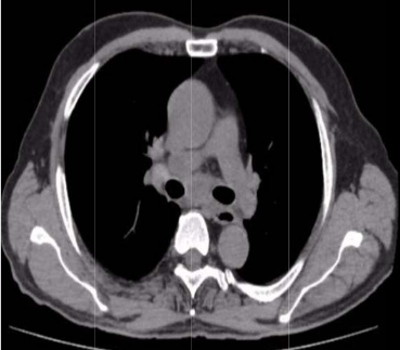
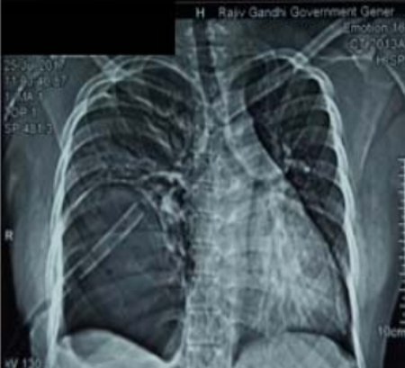
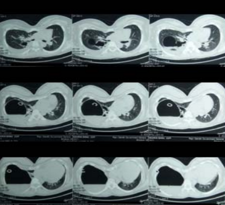
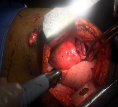
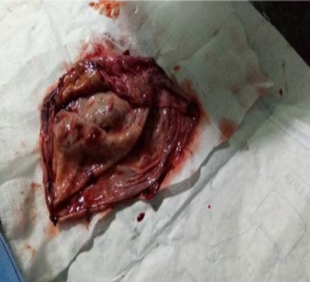
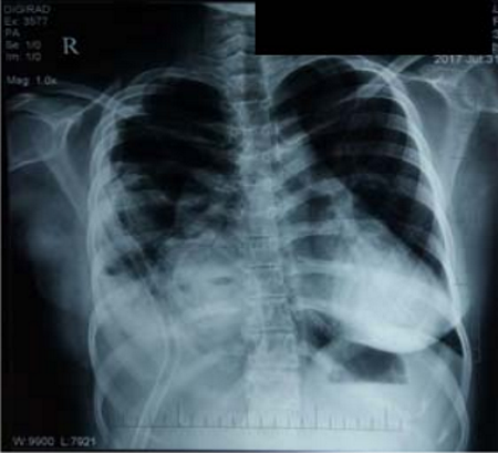
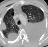
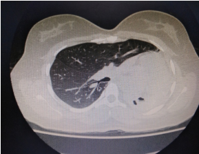
The indications for surgery are the complications and sequelae for tuberculosis. Hemoptysis, destroyed or bronchiectatic lungs, empyema with or without bronchopleural fistula are some of the most common conditions with which patients are referred for surgery.

How to cite this article: Ajay Narasimhan., Editorial, JAPT 2018; 2:47-48

Multi drug resistant tuberculosis is a complex medical entity which forms an important
indication for surgery in a specialised subset of patients. These situations include localised disease, adequate trial of ATT has been given, drug failure and patients are chronically symptomatic.

Surgery for patients with tuberculosis is more difficult than it appears to be. It is of utmost importance that the patient and his relatives are counselled thoroughly regarding the nature of disease and outcome after surgery. They should also be informed about the natural course of disease without any surgical intervention. Pre operative pulmonary rehabilitation plays a major role in improving the pre operative physical fitness of the patient. This includes aggressive chest physiotherapy, postural drainage, nebulisers and antibiotics if necessary. Most of the patients with tuberculosis are nutritionally depleted. Nutritional supplementation plays a major role in improving the post operative outcomes after surgery and reduces the risk of complications such as bronchopleural fistulae and prolonged air leaks.

The outcomes after TB surgery are improved if great detail is given in the post operative period.This includes wound care, care of chest tubes and post operative pulmonary exercises. The common complications after surgery for tuberculosis may be divided into surgical and systemic. The surgical complications include bronchopleural fistula, prolonged air leaks, bleeding and post resectional spaces and empyemas. The medical complications may include respiratory failure, chronic malnutrition.

The advent of minimally invasive thoracic surgical techniques has brought about a renewed zeal in the field of thoracic surgery for tuberculosis. A multidisciplinary team approach involving the pulmonologist, thoracic surgeon, interventional radiologist and infectious disease specialists must work together to actively identify TB patients who are likely to benefit from surgery.

697 views

CT Guided Transthoracic Lung Biopsy – An Experience from a Tertiary Care Centre Hospital

By | volume1-issue2 | No Comments

690 views

Aneeshkumar S et al.: CT Guided Transthoracic Lung Biopsy – An Experience from a Tertiary Care Centre Hospital

Original Article

CT Guided Transthoracic Lung Biopsy – An Experience from a Tertiary Care Centre Hospital

Aneeshkumar.S 1 , Narasimhan.R 2 , Sunder.K 3 and Sundararajan.L 4

1Consultant Pulmonologist, aneesh2907@gmail.com, 2 Senior Consultant Pulmonologist, drnarasimhan@gmail.com
3Senior Consultant Radiologist, sunkap19@yahoo.com, 4 Senior Consultant Pulmonologist, sundar1967@gmail.com
1Department of Respiratory Medicine, KIMS Multispeciality Hospital, Kottiyam, Kollam-691571
2Department of Respiratory Medicine, Apollo Hospitals, Chennai-600006

ABSTRACT:

Background: In 1883, Leyden described percutaneous lung biopsy, but it was not until 1970’s that image guided fine needle chest biopsy gained widespread acceptance. Haaga and Alfidi reported CT-guided thoracic biopsy in 1976. Currently, CT-guided core biopsy is playing an increasing role in the diagnosis of benign as well as malignant diseases, cellular differentiation, somatic mutation analysis, and molecular finger print analysis. Small tissue biopsy obtained through minimally invasive methods have become the primary diagnostic tools for the pathologic characterization and testing of lung masses
Aim: To evaluate the diagnostic accuracy and complications of CT guided transthoracic lung (core) biopsy in radiologically visible lung lesions
Materials and methods: One hundred and one patients who underwent CT guided transthoracic needle aspiration lung biopsy (TNAB) for radiologically visible lung lesions were studied. After obtaining informed consent, the patient was appropriately positioned depending on the site of the lesion. A coaxial 18G biopsy needle (by cook) was used for biopsy. An immediate CT screening was done post procedure to r/o complications.
Results: Transthoracic needle aspiration biopsy was diagnostic in 88% (89/ 101) of patients. In case of  alignancy even the histological type of tumor was known and adequate tissue was present for molecular sampling. The complication rate (pneumothorax) following CT guided biopsy was 5% (5/101)with 1%(1/101)requiring ICD insertion. One patient had minimal haemoptysis which subsided with conservative management.
Conclusion: CT guided lung biopsy is a safe, cost effective and useful procedure for determination of different lung lesions with diagnostic challenge. The complication rates are very few in experienced hands.
Keywords: CT guided transthoracic lung biopsy, diagnostic accuracy, cost effective, complication rate

Corresponding Author: Dr.Aneeshkumar .S, KIMS Kollam Multispeciality Hospital, Sithara junction, Kottiyam P.O., Kollam-691571, India.
Phone number: +91-9497406582 E-mail address : aneesh2907@gmail.com

How to cite this article: Aneeshkumar.S, Narasimhan.R, Sunder.K and Sundararajan.L, CT Guided Transthoracic Lung Biopsy – An Experience from a Tertiary Care Centre Hospital, JAPT 2018; 2:49-52

Introduction

The major challenge when dealing with radiological opacities, especially mass lesion, is to arrive at a conclusive diagnosis. More and more pulmonary lesions, which cannot be found by conventional chest X-rays, are being detected by computed tomography (CT) screening; however, the problem of accurately diagnosing these lesions remains1.

Transthoracic needle aspiration biopsy (TNAB) of the intrathoracic lesions is a well established technique for obtaining tissue for histopathological examination and various other tests 2,3. Haaga and Alfidi reported computed tomography (CT)-guided biopsy in the 17th century, and numerous reports since that time have shown TNAB procedures to be both effective and accurate 4. The diagnostic accuracy has been reported as greater than 80% for benign disease and greater than 90% for malignant disease 5,6.

Materials and methods

This was a prospective observational study conducted at Apollo hospitals, Chennai, from March 2013-September 2015. Patients above the age of 18 years, with radiologically visible lung lesions, undergoing CT guided TNAB were included in the study. Informed consent was obtained from each patient after explaining the risks and benefits. Most of the lesions (around 70%) were peripherally based and few lesions were central (not perilihar). The size of the lesions ranged from around 2.5 cm in case of nodules to 6-7 cm in case of mass lesions. Centrally located lesions which looked accessible via bronchoscope, Fibreoptic bronchoscope was attempted. The patient was taken up for the CT guided TNAB after initial investigations were inconclusive and the lesion posed a diagnostic challenge with malignancy being a high possibility in most of the cases. Patients not willing or cooperating for the procedure or patients with bleeding diathesis, or severe COPD, or contralateral pneumonectomy, or severe PAH were excluded from the study.

Procedure for TNAB

Platelet counts, APTT, PT and INR values were checked before the procedure. Any anticoagulant medication was discontinued 4-5 days to the procedure 7. Fasting for two hours was advised prior to the procedure. Patient was placed in supine, prone or lateral decubitus position depending on the site of lesion. Appropriately placed improvised metal markers were used to determine the optimal cutaneous entry point. The depth of the lesion from the chest wall was noted with the help of a CT scan prior to the procedure. A coaxial 18G biopsy needle (by cook) was introduced through the marked site. Needle direction was planned to have the most vertical approach to the pleura, the shortest distance to the lesion and to avoid penetrating bullae, vessels and fissures. After confirming the position, the stillete was removed and a 20 G needle was inserted and biopsy samples were taken. 4-6 bites were taken depending on the requirement. Size of the biopsy sample (needle throw) was 10 mm for smaller lesions and 20 mm for larger lesions. An immediate screening CT was done post procedure to r/o complications.

Results

One hundred and one patients who underwent CT guided TNAB over a period of two and a half years were assessed. 71/101(70%) were males and 30/101(30%) were females. The mean age group of the patients was 56 ± 13.8. The type of radiological lesions and the lobe wise distribution of the lesions are shown in table 1. The various pathological diagnosis of patients who underwent TNAB are shown in table 2.

Table 1, Showing the type and distribution of radiological lesions

Type of radiological lesions % Lobar distribution of lesions =n(%)
Cavity(n=10) 9.9 Upper(R or L) 43 (42.5)
mass lesion(n=67) 66.3 Upper(R or L) 41 (40.5)
nodules(n=24) 23.7 Middle 5 (5)
Lingula 2 (1.9)
Bilateral 10 (9.9)

Table2.showing the pathological diagnosis of the lesions

Pathological findings %
Adenocarcinoma 38.6
NSCLC 10.5
Chronic inflammation 3.0
HP 0.9
TB 10.9
Organizing Pneumonia 0.9
Small Cell Ca 3.0
Neuroendocrine tumour 2.0
Squamous cell ca 8.9
Aspergilloma 0.9
Sarcoidosis 4.0
Eosinophilic Pneumonia 0.9
Lymphoma 3 4.0
Malignant round cell tumour 0.9
Non diagnostic 11.6

The diagnostic yield following CT guided TNAB was 88% (89/101). In patients diagnosed with malignancy even the histological type was identified and sufficient amount of tissue was available for molecular typing. 5% (5/101) had pneumothorax and 0.9% (1/101) had hemoptysis following TNAB. Out of the 5 patients who developed pneumothorax only 1 required intercostal drainage, while others were managed conservatively by observation and high flow oxygen. Patient who developed haemoptysis was also managed conservatively. The patient who was uneventful post procedure was discharged on the same day of the procedure after observation for four hrs. Patients who developed pneumothorax requiring observation was discharged after 24 hrs and patients who required ICD insertion were discharged after 48 hrs.

Discussion

CT‐guided TNAB allows precise location of target lesions and are cost effective as they shorten the period from admission to diagnosis, decrease the number of surgical procedures and shorten the time of hospital stay as well as require only local anaesthesia 8. The existing biopsy concepts were established in the age of cyto toxic chemotherapy, and were based on the safety, invasion, and cost 9,10.

As the oncology developed into the era of individualized treatment, tumor biopsy aimed to meet not only evidence of malignancy, but also molecular profiling and translational study which is easily achieved with the help of a TNAB. The histological type of malignancy may be sometimes difficult to be determined by a bronchoscopic biopsy, probably due to the small size of the tissue obtained.

Once we encounter a radiological lesion the point of utmost concern should be to decide on the appropriate investigation that is rapid, cost effective, causes minimal complications and gives an accurate diagnosis. With the advances in medical fields various diagnostic modalities are available to obtain a tissue for biopsy, few among which includes bronchoscopic biopsy, CT guided TNAB, Endobronchial ultrasound guided TNAB(EBUS-TNAB), Endoscopic ultrasound guided TNAB(EUS-TNAB) and ultrasound guided biopsy. The quality of a physician rests on selecting an appropriate investigating modality for one’s own patient. As shown in our study the diagnostic yield of TNAB was 88% (89/101) which is very much on par when compared with other studies. In those diagnosed with malignancy even the histological type was known and also there was enough tissue left for molecular sampling. This prevents the unwarranted delay in starting appropriate treatment. Recent studies have even shown the diagnostic efficacy of TNAB even in evaluation of solitary ground glass opacities 11.

The fear of complications, mainly pneumothorax and pulmonary hemorrhage is what restricts a physician from subjecting a patient to TNAB. The rate of pneumothorax after TNAB reported in literatures range from 8% 12 to 61% 13.  Yeow et al. in a study of 660 consecutive biopsies showed that patients with lesions ≤ 20 mm have a higher incidence of pneumothorax than those with larger lesions 14.  Also the pleura to lesion distance influence the risk of pneumothorax with Haramati and Austin reporting a negligible risk of pneumothorax for lesions abutting on the pleura and not requiring the traversal of aerated lung 15. The size of the lesion of the patients in our study ranged from 24 mm in patients with nodule to 6-7 cm, in patients presenting with mass lesion.

The complication rate in our study was only 5% (5/101) with 1 % (1/101) requiring ICD insertion. Since the number of punctures can significantly affect pneumothorax and chest tube insertion rate, investigators have suggested use of a coaxial technique for CT-guided thoracic interventions 14. In our centre also, a coaxial technique was applied and the complication rates encountered are very few. Air embolism is another fatal complication which can occur during the procedure that has to be kept in mind.

Patients who remained undiagnosed (11.6%)) even after TNAB underwent either VATS/open lung biopsy. Though all the patients had a diagnosis post procedure, the hospital stay prolonged to a minimum of 5 more days and also the cost was high. Moreover the procedure was done under general anaesthesia adding to another risk. Because of the minimal invasive nature of TNAB it has widely replaced Open /VATS lung biopsies.

Conclusion

Selecting an appropriate investigating modality which is accurate, safe and cost effective is very much important when dealing with radiological lesions. CT guided TNAB is one such useful diagnostic procedure for determination of different lung lesions which can easily be done under a local anaesthesia. The diagnostic accuracy is very high and complications are few in experienced hands.

REFERENCES

  1. Jemal A, Bray F, Center MM, et al. Global cancer statistics. CA Cancer J Clin 2011;61:69e90.
  2. Klein JS, Salomon G, Stewart EA. Transthoracic needle biopsy with a coaxially placed 20-gauge automated cutting needle: results in 122 patients. Radiology 1996;198:715-720
  3. Manhire A, Charig M, Clelland C, Gleeson F, Miller R, Moss H, et al. Guidelines for radiologically guided lung biopsy. Thorax 2003;58:920-936
  4. Haaga JR, Alfidi RJ. Precise biopsy localization by computed tomography.Radiology 1976;118:603-7.
  5. Tsukada H, Satou T, Iwashima A, Souma T. Diagnostic accuracy of CT-guided automated needle biopsy of lung nodules. Am J Roentgenol 2000;175:239-43.
  6. Westcott JL. Percutaneous transthoracic needle biopsy. Radiology 1988;169:593-601
  7. Manhire A, Charig M, Clelland C, Gleeson F, Miller R, Moss H, et al. Guidelines for radiologically guided lung biopsy. Thorax. 2003;58:920–936.
  8. Zafar N, Mannudim S. Mediastinal Biopsy. Cancer 1995;76:1065-8.
  9. Yung RC. Tissue diagnosis of suspected lung cancer: selecting between bronchoscopy, transthoracic needle aspiration, and resectional biopsy. Respir Care Clin N Am 2003;9:51-76.
  10. Rivera MP, Mehta AC, American College of Chest Physicians. Initial diagnosis of lung cancer: ACCP evidence-based clinical practice guidelines (2nd edition). Chest 2007;132:131S-148S
  11. Yi-Feng Z, Li-Ming J, Wei-Min M, Zhi-Qiang H. Percutaneous computed tomography-guided lung biopsy of solitary nodular ground-glass opacity. J Can Res Ther 2015;11:C231-3.
  12. Westcott JL.Percutaneous transthoracic needle biopsy Radiology 1988;169:593-601
  13. Fink I,Gamsu G,Harter LP.CT guided aspiration biopsy of the thorax.J Comput Assist Tomogr 1982;6:958-962ja
  14. Yeow KM, See LC, Lui KW, Lin MC, Tsao TC, Ng KF, et al. Risk Factors for pneumothorax and bleeding after CT guided percutaneous coaxial cutting needle Biopsy of Lung Lesions. J Vasc Interv Radiol 2001;12:1305‐12.
  15. Haramati LB, Austin JH. Complications after CT‐guided biopsy through aerated versus nonaerated lung. Radiology 1991;181:778.
  16. Laurent F, Michel P, Latrabe V, Tunon de Lara M, Marthan R. Pneumothoraces and chest tube insertion after CT- guided transthoracic lung biopsy using a coaxial technique: incidence and risk factors. AJR Am J Roentgenol 1999; 172: 1049-53.

Abbrevations

TNAB–6 Transthoracic needle aspiration lung biopsy:8-9
ICD– Intercostal drainage
CT– Computed tomography
COPD – Chronic obstructive lung disease
PAH – Pulmonary artery hypertension
APTT – Activated prothromboplastin time
PT – Prothrombin time
INR – International normalized ratio
EBUS – Endobronchial ultrasound
EUS – Endoscopic ultrasound
VATS – Videoassisted thoracic surgery.

616 views

Role of Endobronchial Ultrasound Guided real time Transbronchial Needle Aspiration in Mediastinal Lymphadenopathy: A Case Series

By | volume1-issue2 | No Comments

690 views

Avinash Peddi et al: Role of Endobronchial ultrasound guided real time transbronchila needle aspiration in ….

Original Article

Role of Endobronchial Ultrasound Guided real time Transbronchial Needle Aspiration in Mediastinal Lymphadenopathy: A Case Series

Avinash Peddi 1, Sridhar.R 1 and Narasimhan.R 2

1DNB Post Graduate, Department of Respiratory Medicine, Apollo Main Hospital, Chennai.
2Senior Consultant, Department of Respiratory Medicine, Apollo Main Hospital, Chennai.

ABSTRACT:

Introduction: Mediastinal lymphadenopathy is expected to be the common mediastinal masses because this is caused by high prevalent diseases like tuberculosis, lung cancer metastasis, lymphoma and sarcoidosis etc. Mediastinal lymphadenopathies are difficult to biopsy since they are nearer to major vessels and airways and located commonly in the middle compartment. Hence mediastinal lymphadenopathy is under represented in many studies since unable to obtain tissue diagnosis. The newer techniques EBUS and EUS are made easier to approach the mass like lymphadenopthy in middle mediastinum with less complication.
Objective: To know the effectiveness of real‐time EBUS in the evaluation of mediastinal lymphadenopathy.
Methodology: In our study 50 cases of mediastinal lymphadenopathy diagnosed by CT scan chest which were subjected to EBUS guided real time TBNA of mediastinal nodes. TBNA Specimens were sent for histopathological examinations and reports were collocated and analyzed.
Results: Among 50 cases of mediastinal lymphadenopathy, arrived diagnosis in EBUS TBNA in 42 cases and inconclusive results in other 8 cases. Among 42 cases, tuberculosis in 17 cases [34%], malignancy in 16 cases [32%], sarcoidosis in 8 cases [16%], sub acute inflammation in 1 case [2%].
Conclusion: Before the advent of EBUS, Percutaneous transthoracic needle biopsy/FNAC (under CT or ultrasound guidance) or surgical procedures [VATS or Mediastinoscopy] were considered as the initial method of choice in evaluation of mediastinal lesions. EBUS techniques has demonstrated utility in the diagnosis of mediastinal lymphadenopathy secondary to malignancy, tuberculosis and sarcoidosis and easier and lesser complication than more invasive procedures like VATS or mediastinoscopy.
Keywords: Mediastinal lymphadenopathy, EBUS-TBNA, Tuberculosis, Sarcoidosis.

Corresponding Author: Dr. Avinash Peddi, DNB post graduate, Department of Respiratory Medicine.
Email: dr.sridhar.ily@gmail.com.

How to cite this article: Aneeshkumar.S, Narasimhan.R, Sunder.K and Sundararajan.L, CT Guided Transthoracic Lung Biopsy – An Experience from a Tertiary Care Centre Hospital, JAPT 2018; 2:49-52

Introduction

Mediastinal lymphadenopathy is expected to be the common mediastinal masses because this is caused by high prevalent disease like tuberculosis, lung cancer metastasis, lymphoma and sarcoidosis etc; especially in very high tuberculosis prevalent country like India. Mediastinal lymph node enlargement is most often due to lymphoma 2, metastatic cancer 1, granulomatous inflammation such as that caused by sarcoidosis or infection. Infection should be considered when the adenopathy is associated with a pulmonary opacity. Tuberculosis is a notable cause of mediastinal adenopathy that can mimic sarcoidosis or malignancy and should be suspected in a host with known tuberculosis risk factors, such as recent known exposure or residence in an endemic area 3.

How to cite this article: Avinash Peddi, Sridhar.R and Narasimhan.R, Role of Endobronchial Ultrasound Guided Real Time Transbronchial Needle Aspiration in Mediastinal Lymphadenopathy: A Case Series, JAPT 2018; 2:53-57

Fungal infections that cause granulomas, particularly histoplasmosis, may present with mediastinal and hilar adenopathy in the absence of a pulmonary opacity 4. Numerous less common causes of mediastinal adenopathy are described, including Castleman’s disease or angiofollicular lymphoid hyperplasia.

Tuberculous lymphadenitis is another important consideration for middle mediastinal mass. Tuberculosis (TB) is responsible for up to 43% of all of peripheral lymphadenopathy in the developing world. Total estimated incidence of tuberculous lymphadenopathy was 30.8 per 100 000 population in India. The cervical region is most frequently involved, but mediastinal involvement occurs in approximately 27% of cases 5.

For evaluation of mediastinal adenopathy or other lesions in the middle mediastinum, transbronchial needle aspiration (TBNA) via the fiberoptic bronchoscope offers a less invasive option to surgical mediastinoscopy 6-8. Endobronchial ultrasound-guided transbronchial needle aspiration (EBUS-TBNA) is a recent technology that has significantly improved the ability of pulmonologists to diagnose and stage non–small cell lung cancer in a minimally invasive manner. With the advent of a curvilinear ultrasound probe integrated at the end of the bronchoscope, TBNA with a 22-gauge needle can be performed under real-time ultrasonographic guidance 9-11. Endoscopic biopsies are generally preferred nowadays in view of lesser complications and ease compared to surgical interventions.

Objective

Objective To know the effectiveness of real‐time EBUS in the evaluation of mediastinal lymphadenopathy.

Methodology

In our study 50 cases of mediastinal lymphadenopathy diagnosed by CT scan chest which referred for EBUS-TBNA to department of respiratory medicine, Apollo main hospitals, Chennai were included after obtaining the informed consent. All these patients were subjected to EBUS guided TBNA under general anaesthesia and localized the enlarged mediastinal node in station 4R, 7 or 10 and then EBUS guided real time TBNA of mediastinal nodes was done. TBNA Specimens were sent for histopathological examinations and reports were collocated and analyzed.

Results

In our study 50 patient with mediastinal lymphadenopathy on CT chest was included among these 30 were males and 20 were females with Male to female ratio of 3:1. Mean age group in our study was 50 years with age range from 22 to 75 years.

Among 50 cases of mediastinal lymphadenopathy, arrived diagnosis in EBUS TBNA in 42 cases and in other 8 cases inconclusive results. Pathological reports of EBUS TBNA sample were listed in following table:1.

Table:1 Pathological reports of EBUS TBNA specimens.

Granuloma Malignancy Subacute Non Inflamation Diagnostic
25 16 1 8
50% 32% 2% 16%

In our study population, final diagnosis of mediastinal lymphadenopathies were tuberculosis in 17 cases[34%], malignancy in 16 cases[32%], sarcoidosis in 8 cases[16%], sub acute inflammation in 1 case[2%] and non diagnostic in 8 cases[16%].

Table: 2 Final diagnosis of mediastinal lymphadenopathy

Tuberculosis 17 34%
Sarcoidosis 8 16%
Malignancy 16 32%
Reactive lymphadenitis 1 2%
Non diagnostic 8 16%
TOTAL 50 100%

Discussion

Endobronchial ultrasound – guided transbronchial needle aspiration (EBUS-TBNA) is a minimally invasive technique allowing sampling of mediastinal lymph nodes via fine needle aspiration under direct sonographic visualisation. It has a low rate of morbidity, and has demonstrated utility in the diagnosis of mediastinal lymphadenopathy secondary to malignancy, lymphoma and sarcoidosis.

In Tremblay A et al randomized controlled trial of 50 patients with clinically suspected sarcoidosis due to the presence of mediastinal and hilar adenopathy, the diagnostic yield of EBUS TBNA was superior to blind TBNA, with a sensitivity of 83% and specificity of 100% 12 . In another study Gupta et al randomized control study in 130 patient of sarcoidosis, also stated that the diagnostic yield of EBUS TBNA was superior to blind TBNA 13. In study by Herth FJF et al stated that EBUS-TBNA is a promising new method for sampling mediastinal lymph nodes 14. It appears to permit more and smaller nodes to be sampled than conventional TBNA, and it is safe. In our study we preferred EBUS TBNA over blind TBNA in all our mediastinal lymphadenopathy and the diagnostic yield of EBUS TBNA was 84% in our study.

EBUS with real-time needle aspiration has shown itself to be extremely useful in assessing nodular metastases. In three of the studies reviewed 15,16,17, imaging techniques, fundamentally CT and FDG-PET, which are frequently used as initial staging methods, displayed a lower sensitivity and specificity than that observed for EBUS–TBNA. Unlike imaging techniques, EBUS– TBNA enables the identification and sampling of lymph nodes ≤1 cm in size.

The sensitivity of real-time EBUS–TBNA for assessment of mediastinal and hilar metastasis seems to be equivalent or even superior to that of mediastinoscopy, the reference technique currently used to assess nodular metastases. According to the results of a recent pooled analysis, the sensitivity of mediastinoscopy ranges from 72–89% 18. EBUS– TBNA could replace mediastinoscopy in an important percentage of cases, though, due to the low NPV observed in some studies, surgical techniques cannot be ruled out in negative cases.

The main limitation of EBUS–TBNA is its inability to visualise posterior nodes (stations 5, 7, 8 and 9) 15. EUS–FNA is a complementary technique that enables visualisation of posterior nodes not visualised by EBUS–TBNA but does not allow the visualisation of the anterior mediastinum 15,16

While the efficacy of EBUS-TBNA is now firmly established in the evaluation of lung cancer, there is also an increasing role for the initial evaluation of isolated mediastinal adenopathy due to other conditions such as sarcoidosis, tuberculosis etc. In our study group, EBUS TBNA showed granuloma in 25 cases. Among these granulomatous lymphadenitis based on additional pathological features like caseating necorsis, presence of AFB and clinicoradiological features along with mantoux test results which was non reactive in sarcoidosis patients.

In a prospective trial by Navani N et al of 77 patients with isolated mediastinal adenopathy, a specific diagnosis of sarcoidosis, tuberculosis, lymphoma, or other malignancy was made in 67 of them, thus obviating the need for a more invasive surgical mediastinoscopy19. In our study population, diagnosed malignant mediastinal lymphadenopathy in 16 [32%] cases and also diagnosed mediastinal lymphadenopathy due to tuberculosis in 17 cases [34%] and sarcoidosis in 8 cases [16%]. EBUS technique is helpful in evaluating mediastinal lymphadenopathy which are difficult to approach by image guided percutaneous techniques.

Conclusion

Mediastinal lymphadenopathies are difficult to biopsy since they are nearer to major vessels and airways and located commonly in the middle compartment. Hence mediastinal lymphadenopathy is under represented in many studies since unable to obtain tissue diagnosis. Before the advent of EBUS, Percutaneous transthoracic needle biopsy/FNAC (under CT or ultrasound guidance) or surgical procedures [VATS or Mediastinoscopy] were considered as the initial method of choice in evaluation of mediastinal lesions. EBUS techniques has demonstrated utility in the diagnosis of mediastinal lymphadenopathy secondary to malignancy, tuberculosis and sarcoidosis and easier and lesser complication than more invasive procedures like VATS or mediastinoscopy.

REFERENCES

  1. McLoud TC, Meyer JE. Mediastinal metastases. Radiologic clinics of North America. 1982 Sep;20(3):453.
  2. Filly R, Blank N, Castellino RA. Radiographic distribution of intrathoracic disease in previously untreated patients with Hodgkin’s disease and non-Hodgkin’s lymphoma. Radiology. 1976 Aug;120(2):277-81.
  3. Medford AR, Agrawal S. Single bronchoscope combined endoscopic-endobronchial ultrasound-guided fine-needle aspiration for tuberculous mediastinal nodes. Chest. 2010 Nov 1;138(5):1274.
  4. Wheat LJ, Conces D, Allen SD, Blue-Hnidy D, Loyd J. Pulmonary histoplasmosis syndromes: recognition, diagnosis, and management. InSeminars in respiratory and critical care medicine 2004 Apr (Vol. 25, No. 02, pp. 129- 144). Copyright© 2004 by Thieme Medical Publishers, Inc., 333 Seventh Avenue, New York, NY 10001, USA.
  5. Index TB. Guidelines. Guidelines on extra-pulmonary tuberculosis for India. World Health Organization. 2016.
  6. Shure D, Fedullo PF. The role of transcarinal needle aspiration in the staging of bronchogenic carcinoma. Chest. 1984 Nov 1;86(5):693-6.
  7. Schenk DA, Bower JH, Bryan CL, Currie RB, Spence TH, Duncan CA, Myers DL, Sullivan WT. Transbronchial needle aspiration staging of bronchogenic carcinoma. American Review of Respiratory Disease. 1986 Jul;134(1):146-8.
  8. Rintoul RC, Tournoy KG, El Daly H, Carroll NR, Buttery RC, Van Kralingen K, van Meerbeeck JP, Rabe KF, Annema JT. EBUS-TBNA for the clarification of PET positive intra- thoracic lymph nodes—an international multi-centre experience. Journal of Thoracic Oncology. 2009 Jan 1;4(1):44-8.
  9. Herth FJ, Becker HD, Ernst A. Ultrasound-Guided Transbronchial Needle Aspiration*: An Experience in 242 Patients. Chest. 2003 Feb 1;123(2):604-7.
  10. Yasufuku K, Nakajima T, Motoori K, Sekine Y, Shibuya K, Hiroshima K, Fujisawa T. Comparison of endobronchial ultrasound, positron emission tomography, and CT for lymph node staging of lung cancer. Chest. 2006 Sep 1;130(3):710-8.
  11. Yasufuku K, Nakajima T, Fujiwara T, Chiyo M, Iyoda A, Yoshida S, Suzuki M, Sekine Y, Shibuya K, Yoshino I. Role of endobronchial ultrasound-guided transbronchial needle aspiration in the management of lung cancer. General thoracic and cardiovascular surgery. 2008 Jun 1;56(6):268-76.
  12. Tremblay A, Stather DR, MacEachern P, Khalil M, Field SK. A randomized controlled trial of standard vs endobronchial ultrasonography-guided transbronchial needle aspiration in patients with suspected sarcoidosis. Chest. 2009 Aug 1;136(2):340-6.
  13. Gupta D, Dadhwal DS, Agarwal R, Gupta N, Bal A, Aggarwal AN. Endobronchial ultrasound-guided transbronchial needle aspiration vs conventional transbronchial needle aspiration in the diagnosis of sarcoidosis. Chest. 2014 Sep 1;146(3):547-56.
  14. Herth FJ, Eberhardt R, Vilmann P, Krasnik M, Ernst A. Real-time endobronchial ultrasound guided transbronchial needle aspiration for sampling mediastinal lymph nodes. Thorax. 2006 Sep 1;61(9):795-8.
  15. Yasufuku K, Nakajima T, Motoori K, Sekine Y, Shibuya K, Hiroshima K, Fujisawa T. Comparison of endobronchial ultrasound, positron emission tomography, and CT for lymph node staging of lung cancer. Chest. 2006 Sep 1;130(3):710-8.
  16. Wallace MB, Pascual JM, Raimondo M, Woodward TA, McComb BL, Crook JE, Johnson MM, Al-Haddad MA, Gross SA, Pungpapong S, Hardee JN. Minimally invasive endoscopic staging of suspected lung cancer. Jama. 2008 Feb 6;299(5):540-6.
  17. Bauwens O, Dusart M, Pierard P, Faber J, Prigogine T, Duysinx B, Nguyen B, Paesmans M, Sculier JP, Ninane V. Endobronchial ultrasound and value of PET for prediction of pathological results of mediastinal hot spots in lung cancer patients. Lung cancer. 2008 Sep 1;61(3):356-61.
  18. Toloza EM, Harpole L, Detterbeck F, McCrory DC. Invasive staging of non-small cell lung cancer*: a review of the current evidence. Chest. 2003 Jan 1;123(1):157S-66S.
  19. Navani N, Lawrence DR, Kolvekar S, Hayward M, McAsey D, Kocjan G, Falzon M, Capitanio A, Shaw P, Morris S, Omar RZ. Endobronchial ultrasound–guided transbronchial needle aspiration prevents mediastinoscopies in the diagnosis of isolated mediastinal lymphadenopathy: a prospective trial. American journal of respiratory and critical care medicine. 2012 Aug 1;186(3):255-60.
546 views

Clinical and Inflammatory Phenotypes of Severe Asthma in Adults: A Comprehensive Review

By | volume1-issue2 | No Comments

690 views

Padmavathy Ramadoss: Clinical and Inflammatory Phenotypes of Severe Asthma in Adults: A Comprehensive Review

Review Article

Clinical and Inflammatory Phenotypes of Severe Asthma in Adults: A Comprehensive Review

Padmavathy Ramadoss

Consultant Interventional Pulmonologist and Intensivist,
Department of Pulmonary Medicine, S.K. Hospital, Edapazhinji, Trivandrum, Kerala, India
dr.padmavathyramadoss@gmail.com

ABSTRACT:

Evidence – based medicine is the norm of the day. Ever increasing recognition of marked phenotypic heterogeneity amongst patients with defined ‘severe asthma’ calls for precise, individualised patient-based treatment. Although international research has identified several clinical and inflammatory phenotypes of severe asthma, one of the innumerable hurdles that must be overcome in daily practice, is bridging of the knowledge gap between researchers and Physicians dealing with asthmatic patients in their daily clinical practice. While more enterprise is required to comprehend the relation between these phenotypes and their elemental disease mechanisms and pharmacological treatment responses, in-depth cognizance of these clinical and inflammatory phenotypes provide a good embarkation point for addressing disease heterogeneity in severe asthma in day to day practice. This review brings to the reader, comprehensive information on the clinical and inflammatory asthma phenotypes, their associations and related biomarkers and aims to emphasise the importance of recognition of these phenotypes in order to bring individualised patient management to the bedside.

Corresponding Author: Dr. Padmavathy Ramadoss, Consultant Interventional Pulmonologist and Intensivist, Department of Pulmonary Medicine, S.K. Hospital, Edapazhinji, Trivandrum, Kerala, India. Email: dr.padmavathyramadoss@gmail.com

How to cite this article: Padmavathy Ramadoss, Clinical and Inflammatory Phenotypes of Severe Asthma in Adults: A Comprehensive Review, JAPT 2018; 2:58-63

Introduction

Pulmonary physicians all over the world rely on certain clinical features and patient characteristics to guide them towards diagnosis and treatment of obstructive airway diseases. But with the bountiful armamentaria of treatment options available these days, the overwhelming permutation and combinations of clinical characteristics of asthma makes it an extremely daunting task to further define a specific treatment plan individualised to a patient. Certain terms have been used in order to further simplify things and make medical jargon understood universally. A Phenotype is defined as ‘The composite, observable characteristics of an organism, resulting from interaction between its genetic make-up and environmental influences, which are relatively stable, but not invariable, with time’ [1]. While Endotypes refer to a distinct subgroup of a disease that have a specific pathobiological mechanism. The idea behind identifying specific phenotypes by cluster analysis is that such group of patients will respond better to a specific therapy. Identifying a particular Endotype will aid in selecting a treatment plan that targets a particular pathological pathway of disease and will help in not only a better outcome but also less adverse effects.

Asthma has been variably defined as a ‘heterogeneous disease characterised by chronic airway inflammation. It is defined by history of wheeze, shortness of breath, chest tightness and cough that vary over time and in intensity, together with variable expiratory airflow limitation’[2]. After confirming a diagnosis of asthma and addressing co morbidities, ‘severe asthma’ is that which requires treatment with high dose inhaled glucocorticoids plus a second controller and / or systemic glucocorticoids to prevent asthma from becoming ‘uncontrolled’ or which remains ‘uncontrolled’ despite this therapy [1]. Thus the heterogeneity of asthma with respect to disease severity, age at onset, allergic sensitization, inflammatory mediators, response to treatments, and natural history motivated research based subdivision of asthma into various phenotypes based on underlying pathophysiological mechanisms. This comprehensive review is in no way exhaustive, yet it provides a generalised overview of the main phenotypes of severe asthma.

Why do we need to phenotype asthma?

Although widely accepted guidelines such as the Global Initiative for Asthma (GINA), the National Asthma Education and Prevention Program (NAEPP) and the British Thoracic Society (BTS) [2,4,5] providing guidance on the definitions,
diagnosis and management of asthma are available, it has become a well established norm that patient phenotypic characteristics and disease severity must be taken into consideration while tailoring individualised asthma management plans. Phenotypic characteristics vary from easily recognisable clinical ones to much more complex ones that require a dedicated labs/ specialised centres in order to characterise cellular or biochemical markers in induced sputum or bronchial biopsies which mostly find place in clinical trial settings and in research. An in-depth clinical understanding of phenotypes of asthma that reflects pathophysiologic processes and disease heterogeneity is imperative in order to
successfully manage patients and provide them with appropriate education and life-style advice and to treat based on the phenotype as well as the severity and control of the disease.

Classification Asthma Phenotypes of Severe

Plethora of evidence exists with regards to the phenotypes and sub phenotypes in asthma. Generally, asthma phenotypes can be sub classified based on clinical settings, physiological criteria, treatment response, prognostic factors, and inflammatory milieu and based on underlying pathobiology. While the earlier classification of allergic versus non-allergic asthma still holds good, hypothesis driven and unbiased approaches towards integration of genetics, biology and clinical characteristics have led way to our current understanding of severe asthma phenotypes. This kind of game changing discovery in  clinical phenotyping of severe asthma has been done by The National Heart, Lung and Blood Institute’s (NHBLI) Severe Asthma Research Program (SARP). SARP identified five clusters of adult patients with mild, moderate and severe asthma. They included three groups of mild, moderate and severe early- onset atopic asthma (based on range of lung function, medication use and frequency of exacerbations), a more severe late-onset obese group of primarily older females with moderate FEV1 reductions and frequent oral corticosteroid use, and a later onset but long duration very severe, less atopic group, with less reversible airflow limitation [3]. Woodruff et al, used a molecular approach and found that IL-13 can stimulate the expression of chloride channel, calcium-activated, family member 1 (CLCA1), periostin, serine peptidase inhibitor, clade B (ovalbumin), and serpin family B member 2 (serpinB2), all of which are over expressed in asthmatic patients [22]. Based on inflammatory markers, severe asthma has been classified into TH2 predominant and TH2 poor phenotypes. Another study differentiated severe asthma into clinical and inflammatory phenotypes in children and adults [6]. A further simplification of clinical phenotypes in asthma would divide them into 3 major types: a) asthma with frequent severe exacerbations, (b) asthma with fixed air flow obstruction, (c) asthma with reduced sensitivity or resistance to corticosteroids [7]. Inflammatory phenotypes can be divided into two: a) Persistent severe Eosinophilic asthma, b) Non Eosinophilic severe asthma.

Clinical Phenotypes

A. Exacerbation prone asthma

This phenotype consists of those suffering from frequent severe exacerbations with relatively stable episodes between exacerbations. Characteristics such as age, gender, race, time of onset (Early onset versus Late- onset), asthma related to periods of menstruation or exercise, history of smoking and in association to co morbid conditions form the basis of further sub phenotypes [7].

Prevalence of frequent exacerbations among severe asthmatics has been found to be around 40% [8]. History of frequent exacerbations was found to be the best predictor for further such episodes due to various environmental and genetic factors. On the basis of age of onset 2 phenotypes have been identified: i) early onset (< 12 years of age) and ii) late-onset (> 12 years of age). Early-onset asthmatics with eosinophilia may have a steroid resistance while late-onset asthma without eosinophilia may be a distinct disease [8]. Two main types of asthma exacerbations in terms of acuity of onset described are i) sudden onset and ii) Slow onset. Older asthmatics with acute, severe airflow obstruction, higher frequency of intubation and ventilation yet shorter hospital stay and fewer hospitalisations form a distinct ‘sudden-onset’ phenotypic syndrome. Pathological changes noted with sudden onset exacerbations were presence of low grade inflammation, usually neutrophilic inflammation and bronchospasm in the absence of inflammation. Eosinophilic inflammation, mast cell degranulation, CD8+ lymphocytes, and abundant mucus plugs characterise slow-onset exacerbations [10].

Perimenopausal onset or severe exacerbation of asthma in certain women due to uncertain mechanisms may represent another phenotype [6]. Potentially modifiable risk factors of exacerbation included exposure to cigarette smoke and allergens, obesity, gastroesophageal reflux, sinusitis, psychological factors, and occupational factors [11]. Non modifiable factors identified were viral infections, race/ethnicity, premenstrual asthma, and genetic factors. Suboptimal medical management, failure to comply with medication orders and psychological factors like depression and anxiety were found to be frequently associated with near fatal asthma [12]. Viruses have been implicated in the seasonal variations noted in asthma exacerbations in both adults and children [13]. This may be because of the fact that epithelial cells of asthmatic airways provide a more conducive environment for rhinoviruses to replicate due to the favourable environment of inflammatory allergic process [14]. Genetic factors linking polymorphisms of IL4 gene and its receptor, chitinase and ILD10 genes have all been indentified [15].

B. Asthma with fixed airflow obstruction

Fixed airflow obstruction (FAO) is defined as reduced post bronchodilator ratio of forced expiratory volume in the 1st second (FEV1) to forced vital capacity (FVC) in a patient receiving treatment with high doses of inhaled corticosteroids (Fluticasone 1000 g/d or equivalent) during a stable phase of the disease for at least 4 weeks. Persistent childhood onset asthma and non-allergic, adult onset asthmatics experience a faster decline in lung function and FAO. This holds true in work-place related asthma also, especially secondary to continued exposure to allergic triggers. Risk factors found associated with development FAO include female gender, smoking history, childhood onset asthma, adult- onset non atopic asthma after an infection (Chlamydia pneumonia, Mycoplasma pneumonia), sensitization to Aspergillus fumigatus, especially in patients with associated bronchiectasis [16]. At the microscopic level, FAO in asthma secondary to structural airway wall remodelling, is characterized by increased airway smooth muscle mass and airway wall fibrosis. This phenotype of severe asthma presents with more frequent exacerbations, increased asthma-related mortality and overall mortality [17].

C. Corticosteroid unresponsive asthma

Severe asthmatics can be corticosteroid dependent, refractory or corticosteroid insensitive patients. Steroid dependant patients are those whose asthma control worsens, or develop exacerbations of the disease or who require higher medication levels, on reducing /stopping the dose of steroids. Such patients almost always require larger doses of steroids with resultant problems related to adverse effects of high dose/long term steroids. Corticosteroid insensitivity, which is probably a better term than resistance, is variable and likely has several underlying mechanisms. When FEV1 improves by <15% after 14 days of oral prednisone (40 mg/kg/day) and by >15% after inhalation of salbutamol, asthma is said to be Steroid-resistant [18] . Responsiveness to steroids is related to fraction of inhaled nitric oxide (FENO) levels , sputum eosinophilia, blood eosinophilia, and the results of asthma control tests, whose usefulness varies in adults and children. The various comorbidities associated with corticosteroid insensitivity are obesity, smoking, low vitamin D levels and non-eosinophilic (low- Th2 inflammation) mainly in adults [19]. In adults, a non-eosinophilic phenotype or rather a relatively neutrophil- predominant subgroup of asthma, has been noted, with data from a mild-to-moderate cohort showing relatively poor corticosteroid sensitivity [20]. In depth perception with respect to the mechanisms causing these different types of corticosteroid insensitivity could pave the path to novel treatments such as p38 mitogen-activated protein kinase inhibitors and histone deacetylase-2 recruiters [19].

Inflammatory Phenotypes

Type 2 inflammation is mediated by eosinophils, basophils, mast cells and Th2 lyphocytes and B cells which produced IgE. Significant research work has led to classification of severe asthma based on amount of Type 2 inflammation into TH2 high/ TH2 low phenotypes [25]. This kind of differentiation of asthma is important because it will aid in the selection of therapies based on individual inflammatory phenotypes. In order to do this, the traditional methods airway sampling like bronchoscopy and lavage have paved way for the advent of several non-invasive methods of sputum sampling paved way to easier phenotyping of severe asthma based on inflammatory biomarkers, cellular components of body fluids like sputum, blood and urine, measurement of FENo using breath analyzers and sampling of volatile organic compounds (VOC) using electronic nose or e-nose which is currently under study (21). Two inflammatory phenotypes of severe asthma: a) Persistent severe Eosinophilic asthma, b) Non Eosinophilic severe asthma. Patients with a non-Eosinophilic phenotype can be further split in two inflammatory phenotypes depending on the level of their airway neutrophilic inflammation: i) Neutrophilic ii) Pauci granulocytic asthma

a. Persistent Severe Eosinophilic asthma(PSEA)

Patients with severe asthma and significant eosinophilia in their respiratory airway samples tend to have more severe disease and exacerbations, worse lung functions with significant peripheral airway involvement and remodelling, sinus disease, and a good response to anti- interleukin-5 (IL-5) antibody therapies [23]. No method of estimation of this inflammatory phenotype is ideal. Buhl et al have proposed diagnostic criteria for PSEA [24]. ATS/ ERS guidelines suggest that treatment of severe asthma be guided by clinical criteria and sputum eosinophil counts (performed in centres experienced in this technique) rather than by clinical criteria alone [1]. As IL-5 plays a critical role in eosinophil differentiation, maturation, recruitment, survival and activation in tissues, anti- IL-5 therapies (such as mepolizumab, reslizumab and benralizumab) have been studied and have all established treatment efficacy with no serious safety concerns in clinical trials. Their additional potential benefits include avoidance of adverse events associated with frequent oral steroid use, the rhinosinusitis and nasal polyps associated with upper airway eosinophilia, and atopic dermatitis associated with IL-4/IL-13 activity [24].

b. Non Eosinophilic Severe Asthma

i. Neutrophil predominant severe asthma
A subset of severe, steroid refractory asthmatics, demonstrate neutrophilic- predominant inflammation during exacerbations, both in adults and in some children. Data also seem to suggest that eosinophilic inflammation too can steroid- refractory and perhaps such combined inflammation may prove to be a biomarker of the most severe form of the disease. One study found that nasal polyps and symptoms of gastro- esophageal reflux (GER) were found to be more frequent among severe asthmatics and in one cohort; GER was associated with sputum neutrophilia [26]. Smoking, obesity, obstructive sleep apnea have all have been found in association with neutrophilic dominant inflammatory asthma in various other studies. Airway dysbiosis may be responsible for some aspects of sputum neutrophilia in asthma. Infections with organisms such as Respiratory syncitial virus, Rhinovirus, Aspergillus fumigates, Chlamydia pneumoniae, Streptococcus pneumoniae, Mycoplasma pneumoniae, Haemophilus influenzae, Moraxella catarrhalis and Staphylococcus aureus have all been found to be associated with neutrophil dominant inflammation [27]. Neutrophil rich inflammatory milieu accelerates airway remodelling, rapid decline in lung function and poor response to therapy with corticosteroids. Macrolide therapy maybe used as an alternate therapeutic agent to target pathogens, with ultimate reduction in sputum neutrophil numbers and improvement in symptoms. Yet, it cannot be considered a viable long-term option because of the potential for inducing macrolide- resistant organisms and risk of adverse cardiovascular outcomes [27].

ii. Severe Paucigranulocytic Asthma (SPGA)
This phenotype of severe asthma is characterized by no evidence of increased sputum or blood eosinophils or neutrophils, with normal levels of the proteolytic enzymes MMP-9 and neutrophil elastase in sputum, and ineffectiveness of anti-inflammatory therapies in controlling symptoms. SPGA represents perhaps an uncoupling of airway obstruction from airway inflammation driven by structural changes within the airways, such as airway smooth muscle hypertrophy. Due to the paucity of inflammatory cells, one may postulate that SPGA may represent a milder form of asthma, Yet a substantial proportion of patients with SPGA (21.7%) were characterized as having severe refractory asthma, and 14.8% of patients with PGA had an Asthma Control Test score of less than 19, suggesting that this subpopulation of PGA is not well controlled despite the absence of inflammatory cells in their nsputum [28].

Conclusion

The clinical, inflammatory and molecular level heterogeneity of severe asthma as a syndrome, has evolved insight-driven diagnostic and management approaches to redefine how we tailor individualised severe asthma management plans. An in-depth knowledge and updated awareness about the newer advances in diagnostic strategies and medical and non-pharmacological management options will help to re-define precision medicine in severe asthma patients.

REFERENCES

  1. Chung KF et al. International ERS/ATS guidelines on definition, evaluation and treatment of severe asthma. Eur Respir J 2014; 43:343.
  2. From the Global Strategy for Asthma Management and Prevention, Global Initiative for Asthma (GINA) 2018. http:// www.ginasthma.org/
  3. Moore WC et al. Identification of asthma phenotypes using cluster analysis in the Severe Asthma Research Program. Am J Respir Crit Care Med 2010; 181:315 -323.
  4. Expert Panel Report 3 (EPR-3): Guidelines for the Diagnosis and Management of Asthma-Summary Report 2007. J Allergy Clin Immunol 2007; 120: Suppl 1, S94–S138.
  5. British guideline on the management of asthma. Thorax 2014; 69: 1–192.
  6. Gaga M, Zervas E, Gibson PG. Allergic and non-allergic factors in severe asthma. Eur Respir Mon. 2011; 51:107-19
  7. Bel E, ten Brinke A . A rational approach to the management of severe refractory asthma. Treat Respir Med 2005;4:365–79.
  8. Wenzel SE, Busse WW. Severe asthma: lessons from the Severe Asthma Research Program. J Allergy Clin Immunol2007;119: 14–21.
  9. Miranda C, Busacker A, Balzar S, Trudeau J, Wenzel SE. Distinguishing severe asthma phenotypes: role of age of onset and eosinophilic inflammation. J Allergy Clin Immunol. 2004; 113:101-108.
  10. Campo P, Rodríguez F, Sánchez-García S, Barranco P, Quirce S, Pérez-Francés C, Gómez-Torrijos E, Cárdenas R, Olaguibel JM, Delgado J; Severe Asthma Workgroup; SEAIC Asthma Committee. Phenotypes and endotypes of uncontrolled severe asthma: new treatments. J Investig Allergol Clin Immunol. 2013; 23(2):76-88; quiz 1 p. follow 88.
  11. Barranco P, Pérez-Francés C, Quirce S, Gómez-Torrijos E, Cárdenas R, Sánchez-García S, Rodríguez-Rodríguez F, Campo P, Olaguibel JM, Delgado J (Severe Asthma working group of SEAIC). Consensus document on the diagnosis of severe uncontrolled asthma. J Investig Allergol Clin Immunol.2012;22:460-75.
  12. Romagnoli M, Caramori G, Braccioni F, Ravenna F, Barreiro E, Siafakas NM, Vignola AM, Chanez P, Fabbri LM, Papi A; and the ENFUMOSA Study Group. Near-fatal asthma phenotype in the ENFUMOSA Cohort. Clin Exp Allergy. 2007; 37:552-7.
  13. Wenzel SE. Asthma exacerbations in relation to severe asthma. Eur Respir Mon. 2011;51: 120-9.
  14. Zambrano JC, Carper HT, Rakes GP, Patrie J, Murphy DD, Platts-Mills TA, Hayden FG, Gwaltney JM Jr, Hatley TK, Owens AM, Heymann PW. Experimental rhinovirus infection challenges in adults with mild asthma: response to infection in relation to IgE. J Allergy Clin Immunol. 2003; 111:1008-16.
  15. Sandford AJ, Chagani T, Zhu S, Weir TD, Bai TR, Spinelli JJ, Fitzgerald JM, Behbehani NA, Tan WC, Paré PD. Polymorphisms in the IL4, IL4RA and FCERIB genes and asthma severity. J Allergy Clin Immunol. 2000; 106:135-40.
  16. Bel EH, ten Brinke A, Sorkness RL. Fixed airway obstruction in severe asthma. Eur Respir Mon. 2011;51:147- 59.
  17. Yii AC, Tan GL, Tan KL, Lapperre TS, Koh MS. Fixed airways obstruction among patients with severe asthma: findings from the Singapore General Hospital-Severe Asthma Phenotype Study. BMC Pulm Med. 2014;14:191. doi: 10.1186/1471-2466-14-191.
  18. Wenzel S. Severe asthma in adults. Am J Respir Crit Care Med.2005; 172:149-60.
  19. Hew M, Chung KF. Corticosteroid insensitivity in severe asthma: significance, mechanisms and aetiology. Internal Med J 2010; 40:323–334.
  20. McGrath KW, Icitovic N, Boushey HA, et al. A large subgroup of mild-to moderate asthma is persistently noneosinophilic. Am J Respir Crit Care Med 2012; 185:612– 619.
  21. Gibson PG, Wang F, He XY, Brightling CE. Noninvasive assessment of inflammation in severe asthma. Eur Resp Mon. 2011; 51:208-17.
  22. Woodruff PG, Boushey HA, Dolganov GM, Barker CS, Yang YH, Donnelly S, et al. Genome-wide profiling identifies epithelial cell genes associated with asthma and with treatment response to corticosteroids. Proc Natl Acad Sci U S A (2007)
    104:15858–63.10.1073/pnas.0707413104.
  23. Haldar P, Brightling CE, Hargadon B, Gupta S, Monteiro W, Sousa A, Marshall RP, Bradding P, Green RH, Wardlaw AJ, Pavord ID. Mepolizumab and exacerbations of refractory eosinophilic asthma. N Engl J Med. 2009; 360:973-84.
  24. Buhl R, Humbert M, Bjermer L, et al. Severe eosinophilic asthma: a roadmap to consensus. Eur Respir J 2017; 49: 1700634 [https://doi.org/10.1183/13993003.00634-2017].
  25. Woodruff PG, et al. T-helper type 2-driven inflammation defines major subphenotypes of asthma. Am J Respir Crit Care Med. 2009;180(5):388–95.
  26. Simpson JL, et al. Neutrophilic asthma is characterised by increased rhinosinusitis with sleep disturbance and GERD. Asian Pac J Allergy Immunol. 2014;32(1):66–74.
  27. Ray A, Kolls JK. Neutrophilic inflammation in asthma and association with disease severity. Trends Immunol. 2017;38(12):942–954
  28. Tliba O, Panettieri RA Jr. Paucigranulocytic asthma: uncoupling of airway obstruction from inflammation. J Allergy Clin Immunol. 2018 Jun 19. pii: S00916749(18)30861–3.
    https://www.ncbi.nlm.nih.gov/pubmed/29928921.
591 views

Role of mi-RNAs in Respiratory Diseases

By | volume1-issue2 | No Comments

690 views

Vimal Raj R et al.: Role of mi-RNAs in respiratory diseases

Review Article

Role of mi-RNAs in Respiratory Diseases

Vimal Raj R1 and Pajanivel R 2

1Assistant Professor, 2Professor & Head,
Department of Pulmonary Medicine, Mahatma Gandhi Medical College & Research Institute, Sri Balaji Vidyapeeth, Puducherry.

ABSTRACT:

Ever since the initial discovery in the early 1990s, microRNAs have become the focus of a multitude of conditions from basic biology to clinical applications in both diagnostic and prognostic strata. Previously they were believed to be of no biological relevance. Even in diseases of the lung, microRNAs have been analyzed in the pathophysiology, as drivers of disease, potential treatment targets, and serum biomarkers; however, much is yet to be understood about these non-coding RNAs for us to fully realize their potential therapeutic use. Here, we give a brief insight into their role in major respiratory diseases unearthed so far and the road ahead to better understand this potential biomarker.
Keywords: miRNA, biomarker, polymerase chain reaction

Corresponding Author: Dr. Vimal Raj R, Assistant Professor, Department of Pulmonary Medicine, Mahatma Gandhi Medical College & Research Institute, Sri Balaji Vidyapeeth, Puducherry.

How to cite this article: How to cite this article: Vimal Raj R and Pajanivel R, Role of mi-RNAs in Respiratory Diseases, JAPT 2018; 2:64-67

What are miRNAs?

Genomic studies revealed that numerous portions of the human genome do not encode conventional protein-coding genes but encode biologically active non-coding RNA species. With the rapid expansion of small RNA interference techniques over the past decade, it is now clear that many small RNA molecules could regulate gene and protein expression.

One class of such small non-coding RNAs is microRNAs (miRNAs), a group of regulatory RNAs of 19–22 nucleotides involved in control of gene expression at the post transcriptional level (1) thereby acting as RNA interfering (RNAi) molecule. While a well-known RNAi molecule, small interfering RNA (siRNA), is a small RNA that is artificially synthesized miRNA exists endogenously in the cell. miRNAs were first discovered in 1993 while studying Caenorhabditis elegans. After seven years (in 2000), let-7, the second miRNA was discovered, again in the C. elegans. In last decade, significant advances have been made in miRNA research leading to the discovery of more than 1,900 miRNAs that have been fully characterized (as per miRBase database viewed in Oct 2018).

Tuberculosis

miRNA spectrum in body fluids can reflect altered physiological and/or pathological conditions. Recent studies have shown that miRNAs are stably present in sputum (3,4) and unique miRNA signatures in sputum are altered in many lung diseases, such as lung cancer and chronic obstructive pulmonary disease (5,6).

Genome wide miRNA expression in sputum supernatant of patients with active pulmonary tuberculosis was also delineated in recent past (10). In this study by Yi Z et al, a total of 95 miRNAs were found to be expressed differentially by microarray and miR-3179, miR-147 overexpressed and miR-19b-2* suppressed in TB patient group compared with controls as observed in the validation cohort by real time polymerase chain reaction (RT-PCR).

Lv et al (7) showed that sputum and serum miRNA-144 expression levels showed significant response to treatment (decreased) after the initiation of treatment (one month after initiation of treatment). Their study involved 3 groups of patients and a fourth- control group. This study also showed a difference in the expression levels between sputum smear positive and smear negative patients (more in smear positive patients). These results indicate that sputum and serum miR-144 could be used as diagnostic indices for TB and even for TB severity. This suggestion is further rationalized by the ROC curve results, which showed both sputum and serum miR-144 to have relatively high sensitivity and specificity for the diagnosis of TB.

miR-144 was one of the microRNAs that were over expressed in active TB patients. Real-time RT- PCR analysis showed that miR-144 was mainly expressed in T cells. Transfection of T cells with miR-144 precursor demonstrated that miR-144 could inhibit TNF-α and IFN-γ production and T cell proliferation. miR-144 has been shown to directly inhibit the expression of autophagy-related gene Atg4a and to participate in the regulation of the autophagy process in M. tuberculosis infection (1). It is concluded that miR-144 might involve in regulation of anti-TB immunity through modification of cytokine production and cell proliferation of T cells.

COPD

Few studies to date have examined the role for microRNAs in COPD. The majority of studies have focused on global patterns of microRNA expression. In two early studies, investigators evaluated the effects of cigarette smoke on microRNA expression in animal models. In the first study, rats were exposed to environmental cigarette smoke versus ambient air control over 4 weeks. Twenty-four microRNAs were significantly down-regulated between smoke-exposed and sham groups (10). In the second study, investigators evaluated the effects of smoke exposure on microRNA expression in mice. The majority of deregulated microRNAs in this study were also down-regulated (11).

Christenson and colleagues conducted a comprehensive analysis for microRNAs and mRNAs within different regions of the lung (12). They profiled eight separate regions within the lungs of eight individuals (six with COPD and two control subjects). Using this strategy, the authors were able to not only identify select deregulated microRNAs but also build and enrich for specific biological pathways. They identified 63 microRNAs that were deregulated in regions of emphysema. Of note, three of the deregulated microRNAs (miR- 638, miR-30c, and miR-181d) had an inverse relationship in expression to several of their predicted targets.

Muscle-specific miRNAs play a relevant role in the regulation of muscle development and repair after injury by targeting different pathways. In COPD, the inspiratory loads to which the respiratory muscle is continuously exposed may be a major player accounting for specific pattern of miRNA expression [13]. In the main inspiratory muscle (diaphragm) of patients with mild-to- moderate and severe COPD, expression of some muscle-specific miRNAs miR-1, miR-133, and miR- 206 is down-regulated. However, the expression of miR-486, miR-27a, miR-29b, and miR-181a does not differ between the patients and the controls [13].

Asthma

The inflammatory response is central to the progression and pathology of asthma and is manifested by a production of IgE and consistent recruitment of leukocytes, in particular eosinophils, together with Th2 cells and mast cells (14). MiR-21 is a microRNA that may be central to this process. Up-regulated in individuals with asthma, miR-21 targets IL-12, a potent cytokine responsible for Th1 cell activation.

Various asthma models were documented to have different expression of miRNAs. Wang et al. identified different expression of miR-145-5p, miR- 636, miR-338-3p, miR-4485, miR-1229-3p, miR-4707- 3p, and miR-3620-3p in the serum of patients with asthma, compared to patients with COPD [20]. A few studies demonstrated different expression of 11 miRNAs in the exhaled breath condensate (EBC) from patients with asthma compared to healthy individuals [21].

Targeting microRNAs may represent a novel potential therapeutic strategy in the treatment of asthma. By inhibiting expression of various microRNAs, such as miR-21, -106a, -126, -145, -155, and -221, abnormal cytokine expression and inflammation can potentially be mitigated.

Lung malignancy

MicroRNAs have been well studied in lung cancer with a multitude of cell, animal, and human studies demonstrating deregulation in lung tumors compared with uninvolved lung tissues (18). Specific microRNAs, although not lung cancer– specific, including let-7, miR-21, miR-29, miR-126, miR-155, and miR-17- 92, appear to be fundamental in tumor biology and thus ideal candidates as biomarkers in solid tumors (19-22). Sozzi and colleagues conducted an independent study demonstrating that a distinct panel of circulating microRNAs could be applied as diagnostic and prognostic biomarkers in lung cancer (17).

Conclusion

The role of microRNAs will be unearthed more and more in the years to come. Research outputs till date show that they will improve the diagnostic and prognostic accuracy of not only respiratory diseases but the entire disease conundrum. It is for us to understand their complex role in human physiology and disease processes. As far as pulmonologists are concerned, the role of individual microRNAs/ a panel of microRNAs in core respiratory diseases need to be analyzed in greater detail to make their usage in mainstream clinical practice a reality in the near future.

REFERENCES

  1. Bartel DP: MicroRNAs: genomics, biogenesis, mechanism, and function. Cell. 2004, 116 (2): 281-297. 10.1016/S0092- 8674(04)00045-5.
  2. Zhou L, Guo L, Tang J, Zhang A, Liu X, Xu G. [miR-144 regulates BCG- and rapamycin-induced autophagy by targeting Atg4a in RAW264.7 cells]. Xi Bao Yu Fen Zi Mian Yi Xue Za Zhi 2015;31:163–7.
  3. Xie Y, Todd NW, Liu Z, Zhan M, Fang H, et al. (2010) Altered miRNA expression in sputum for diagnosis of non-small cell lung cancer. Lung Cancer 67: 170–176.
  4. Yu L, Todd NW, Xing L, Xie Y, Zhang H, et al. (2010) Early detection of lung adenocarcinoma in sputum by a panel of microRNA markers. Int J Cancer 127: 2870–2878.
  5. Oglesby IK, McElvaney NG, Greene CM (2010) MicroRNAs in inflammatory lung disease–master regulators or target practice? Respir Res 11: 148.
  6. Pottelberge GR, Mestdagh P, Bracke KR, Thas O, Durme YM, et al. (2011) MicroRNA expression in induced sputum of smokers and patients with chronic obstructive pulmonary disease. Am J Respir Crit Care Med 183: 898–906.
  7. Yan Lv, Shuai Guo, Xue-Gang Li, Jing-Yu Chi, Yi-Qing Qua, Hai-Lai Zhong. Sputum and serum microRNA-144 levels in patients with tuberculosis before and after treatment. International Journal of Infectious Diseases 43 (2016) 68–73.
  8. Yi Z, Fu Y, Ji R, Li R, Guan Z: Altered micro RNA signatures in sputum of patients with active pulmonary tuberculosis. PLoS One. 2012, 7 (8): e43184-10.1371/journal.pone.0043184.
  9. Yanaihara N, Caplen N, Bowman E, Seike M, Kumamoto K, Yi M, Stephens RM, Okamoto A, Yokota J, Tanaka T, et al. Unique microRNA molecular profiles in lung cancer diagnosis and prognosis. Cancer Cell 2006;9:189–198.
  10. Izzotti A, Calin GA, Arrigo P, Steele VE, Croce CM, De Flora S. Down regulation of microRNA expression in the lungs of rats exposed to cigarette smoke. FASEB J 2009;23:806–812.
  11. Izzotti A, Calin GA, Steele VE, Croce CM, De Flora S. Relationships of microRNA expression in mouse lung with age and exposure to cigarette smoke and light. FASEB J 2009;23:3243–3250.
  12. Christenson SA, Brandsma CA, Campbell JD, Knight DA, Pechkovsky DV, Hogg JC, Timens W, Postma DS, Lenburg M, Spira A. miR-638 regulates gene expression networks associated with emphysematous lung destruction. Genome Med 2013;5:114.
  13. E. Puig-Vilanova, R. Aguil ́o, A. Rodr ́ıguez-Fuster, J. Mart ́ınez-Llorens, J. Gea, and E. Barreiro, “Epigenetic mechanisms in respiratory muscle dysfunction of patients with chronic obstructive pulmonary disease,” PloS ONE, vol. 9, no. 11,Article ID e111514, 2014.
  14. Frieri M. Advances in the understanding of allergic asthma. Allergy Asthma Proc 2007;28:614–619.
  15. M. Wang, Y. Huang, Z. Liang et al., “Plasma miRNAs might be promising biomarkers of chronic obstructive pulmonary disease,”The Clinical Respiratory Journal, vol. 10, no. 1, pp. 104– 111, 2016.
  16. A. Sinha, A. K. Yadav, S. Chakraborty et al., “Exosome- enclosed microRNAs in exhaled breath hold potential for biomarker discovery in patients with pulmonary diseases,” The Journal of Allergy and Clinical Immunology, vol. 132, no. 1, pp. 219–222.e7,2013.
  17. Yanaihara N, Caplen N, Bowman E, Seike M, KumamotoK, Yi M, Stephens RM, Okamoto A, Yokota J, Tanaka T, etal. Unique microRNA molecular profiles in lung cancer diagnosis and prognosis. Cancer Cell 2006; 9:189–198.
  18. Takamizawa J, Konishi H, Yanagisawa K, Tomida S, OsadaH, EndohH, Harano T, Yatabe Y, Nagino M, Nimura Y, etal. Reduced expression of the let-7 microRNAs in human lung cancers in association with shortened postoperative survival. Cancer Res 2004; 64:3753–3756.
  19. Seike M, Goto A, Okano T, Bowman ED, Schetter AJ, Horikawa I, Mathe EA, Jen J, Yang P, Sugimura H, et al. MiR-21 is an EGFRregulated anti-apoptotic factor in lung cancer in never-smokers. Proc Natl Acad Sci USA 2009;106:12085–12090.
  20. Fabbri M, Garzon R, Cimmino A, Liu Z, Zanesi N, Callegari E, Liu S, Alder H, Costinean S, Fernandez-Cymering C, et al. MicroRNA-29 family reverts aberrant methylation in lung cancer by targeting DNA methyltransferases 3A and 3B. Proc Natl Acad Sci USA 2007;104: 15805–15810.
  21. Vosa U, Vooder T, Kolde R, Vilo J, Metspalu A, Annilo T. Meta-analysis of microRNA expression in lung cancer. Int J Cancer 2013;132: 2884–2893.
539 views

Melioidosis Presenting as Isolated Mediastinal Lymphadenopathy

By | volume1-issue2 | No Comments

690 views

R. Gogulakrishnan et al: Melioidosis presenting as isolated mediastinal lymphadenopathy

Case Report

Melioidosis Presenting as Isolated Mediastinal Lymphadenopathy

Gogulakrishnan.R 1, Jayakumar.S 1, Subramony.H 2 and Narasimhan.R 3

1Post graduate, Department of Respiratory Medicine. Apollo Hospitals, Chennai.
2Senior Consultant, Department of General Medicine, Apollo Hospitals, Chennai.
3Senior Consultant, Department of Respiratory Medicine, Apollo Hospitals, Chennai.

ABSTRACT:

Melioidosis has varied clinical presentation involving different organs with pneumonia as common pulmonary presentation. Isolated mediastinal lymphadenopathy as presentation is rare. Herein, we describe a case of Melioidosis identified by endobronchial ultrasound guided transbronchial needle aspiration of subcarinal lymph node. This case stresses the need for clinical suspicion of melioidosis in patients from endemic areas and the role of endobronchial ultrasound in diagnosing it.
Keywords: Melioidosis, Mediastinal lymphadenopathy, EBUS-TBNA

Corresponding Author:Dr. R. Gogulakrishnan, Post graduate, Department of Respiratory Medicine. Apollo Hospitals, Chennai-29.

How to cite this article: Aneeshkumar.S, Narasimhan.R, Sunder.K and Sundararajan.L, CT Guided Transthoracic Lung Biopsy – An Experience from a Tertiary Care Centre Hospital, JAPT 2018; 2:49-52

Introduction

Melioidosis is known as “the great mimicker” due to similarity in presentation with other diseases and difficulty in diagnosis. [1] Pneumonia is the most common presentation of melioidosis accounting for 51% of cases. Mediastinal lymphadenopathy was present in only 3% of cases, of which 70% of cases were associated with pneumonia. [2] Our search yielded less than 10 cases of isolated mediastinal lymphadenopathy. [3][4][5]Only 2 cases were diagnosed as melioidosis by endobronchial ultrasound guided transbronchial needle aspirate. [6][7]

Case History

A 62-year-old male from north east India came with complaints of persistent high grade fever and dry cough for the past 3 months. He is a retired teacher by occupation and now does paddy farming. There is history of loss of appetite and no history of loss of weight. He was a non-smoker and did not had any prior pulmonary problems. He was a known hypertensive and not a diabetic. Examination was unremarkable. His oxygen saturation was 95% on room air. His basic blood investigations were within normal limits with a white blood cell count of 8,370 cells/cu.mm. HIV serology was negative. His chest radiograph showed normal lung parenchyma with widening of subcarinal angle suggesting subcarinal lymphadenopathy. High resolution computed tomography revealed subcarinal lymphadenopathy with areas of hypoattenuation suggesting necrosis [Figure 1]. Lung parenchyma did not show any infiltrates.

Endobronchial ultrasound guided transbronchial needle aspiration was done which aspirated purulent material from the subcarinal lymph node [Figure 2]. Post procedure patient was s stable and was shifted toward. He was continued on empirical antibiotics ( Cefoperazone+sulbactam). He developed Ventricular tachycardia and Ventricular fibrillation and was reverted to sinus rhythm with Amiodarone and electric calc cardioversion n. Later, he developed cardiac are est a succumb and bed to his illness.

His transbronchial needle aspirate histology was suggestive of subacute inflammation. Acid fast bacilli stain and Gene Xpert were negative. Bacterial culture of lymph node aspirate grew Burkholderia pseudomallei.

Figure(1) – Computed tomography of chest showing subcarinal lymphadenopathy.

Figure(2) – EBUS guided TBNA being performed in subcarinallymp node

Discussion

Melioidosisis caused by gram negative bacteria burkholderia pseudomallei present in soil. it is endemic in south east asia and nothern part of Australia. Disease can be acquired by inhalation, inoculation or ingestion. [8] Mediastinal lymphadenopathy can occur following either inhalation or hematogenous spread secondary to inoculation.

Chronic renal disease, chronic lung disease and age more than 45 years were considered as risk factors for melioidosis. [9] Apart from age our patient did not have any other risk factors. Melioidosis in its chronic form can mimic as malignancy or tuberculosis.

Aspiration of purulent material from EBUSTBNA increased our suspicion to infectious aetiology, however a malignant cause could not be ruled out. Microbiological culture in the background of high clinical suspicion for infection from lymph node aspirate has increased the diagnostic yield.[10]

Conclusion

Melioidosis has varied clinical presentations. Hence, a high index of clinical suspicion for melioidosis in patients coming from EBUS-TBNA samples can help in early diagnosis of melioidosis.

REFERENCES

  1. Yee KC, Lee MK, Chua CT, Puthucheary SD. Melioidosi is, the great mimicker: a report of 10 cases from Malaysia. J Trop Med Hyg. 1988;91:249 9-54.
  2. Currie BJ, Ward L, Cheng AC. The epidemiology and clinical specctrum of melioidosis: 540 cases from the 20 ye ear Darwin pros spective study. PLoSNegl Trop Dis. 2010;4:e900.
  3. Chlebicki M, Tan B. Six causes of suppurative lymphadenits caused by Burkholderia pseudomallei infection. Transactions of the Royal Society of Tropical Medicine and Hygiene. 2006;100:798-801.
  4. Saravu K, Mukhopadhyay C, Eshwara VK, Shastry BA, Ramamoorthy K, Krishna s, et al. Melioidosis presenting with mediastinal lymphadenopathy masquerading as malignancy:a case report. J Med Case Rep. 2012;6;28.
  5. Saravu K, Vishwanath S, Kumar RS, Barkur AS, Varghese GK, Mukhopadhyay C, et al. Melioisdosis-1 case series from south india. Trans R Soc Trop Med Hyg. 2008;102 Suppl:S18-20.
  6. ChanHP, Yip HS. Mediastinal Lymphadenopathy:Melioidosis Mimicking Tuberculosis. Tropical Medicine and Health 2015;43:93-94.
  7. S.Roche, R.J.Fahy, A.Mclaughlin, J.M.Keane. Melioidosis- a rare case of Mediastinal Lymphadenopathy. Am J Respir Crit Care Med. 2018;197:A5450.
  8. White NJ. Melioidosis. Lancet 2003 May 17;361:1715-22.
  9. Currie BJ, Jacups SP, Cheng AC, Fisher DA, Anstey NM, Huffam SE. et al. Melioidosis epidemiology and risk factors from a prospective whole-population study in northern Australia. Trop Med Int Health. 2004;9:1167-74.
  10. Harris RM, Arnaout R, Koziel H, Folch E, Majid A, Kirby JE. Utility of microbiological testing of thoracic lymph nodes sampled by endobronchial ultrasound-guided transbronchial needle aspiration (EBUS-TBNA) in patients with mediastinal lymphadenopathy. DiagnMicrobiol Infect Dis.2016;84:170-4.
506 views

Post-Partum Presentation of Ruptured Bronchogenic Cyst

By | volume1-issue2 | No Comments

690 views

Ajay Narasimhan et al.: Post-partum presentation of Ruptured Bronchogenic cyst

Case Report

Post-Partum Presentation of Ruptured Bronchogenic Cyst

Ajay Narasimhan 1, Jayagovardhanan Govindasamy Dayalu 2, Dinesh Shanmugasundaram 3 and Arvind Venkatasamy 4

1Assistant Professor, 2 Assistant Professor, 3 Associate Professor, 4 Post Graduate
Institute of Cardiothoracic Surgery, Rajiv Gandhi Government General Hospital, Madras Medical College, Chennai, Tamil Nadu, India

ABSTRACT:

Mediastinal cysts account for 20% of all mediastinal masses. A 22-year-old pregnant female was admitted for delivery. She developed dyspnea on the 3rd day of delivery. Chest X-ray revealed a large right hydropneumothorax and tube thoracostomy was done. She was referred to us for further management. We decided to operate. At surgery, a ruptured cystic lesion was seen in the posterior mediastinum separate from all three lobes. It was communicating with the bronchus. The cyst was excised completely and bronchial communication was sutured. The lung expanded completely. Post operative period was uneventful. Histopathology revealed it to be a bronchogenic cyst.

Corresponding Author: Dr. Ajay Narasimhan M.Ch, Assistant Professor, Institute of Cardiothoracic Surgery, Rajiv Gandhi Govt. General Hospital, Madras Medical College, EVR Periyar Salai, Chennai – 600003. Ph : +91 9940137747

How to cite this article: Aneeshkumar.S, Narasimhan.R, Sunder.K and Sundararajan.L, CT Guided Transthoracic Lung Biopsy – An Experience from a Tertiary Care Centre Hospital, JAPT 2018; 2:49-52

Introduction

Mediastinal masses are uncommon conditions that pose a diagnostic and therapeutic challenge to thoracic surgeons. Mediastinal cysts account for 20% of all such cases. A variety of names have been attributed to mediastinal cysts such as bronchogenic, bronchoesophageal, enterogenous, esophageal, duplication cysts. Fallon and associates in 1954 utilised the cysts embryological origin and anatomical location to classify these lesions. Most of the mediastinal cysts are congenital in origin and embryologic development is an important factor in classification. (1)

Case report

A 22 year old female was admitted at term for delivery. She had a vaginal delivery which was uneventful. She delivered a healthy female baby. She was due to be discharged from the hospital on the 3rd day when she developed breathlessness. A Chest X-ray was taken which showed right sided hydropneumothorax for which tube thoracostomy was done.

How to cite this article: Ajay Narasimhan, Jayagovardhanan Govindasamy Dayalu, Dinesh Shanmugasundaram and Arvind Venkatasamy, Post-Partum Presentation of Ruptured Bronchogenic Cyst, JAPT 2018; 2:71-73

She drained around 1.5 litres of purulent looking fluid and had massive air leak. She was then referred to our department for further management. Her blood investigations were within normal limits. We ordered for a computerised tomography of the chest.

It showed a large cystic lesion within the right lower lobe of the lung with an air fluid level and intercostal tube in situ. Bronchoscopy was unremarkable. At this stage we had a differential diagnosis of a ruptured lung abscess, lung cyst or a hydropneumothorax. In view of persistent drainage and air leak, we decided to go ahead with surgery. We informed the patient that she might need a right lower lobectomy.

After pre anaesthetic evaluation, she was taken up for surgery. Under general anaesthesia with a double lumen endotracheal tube, a posterolateral thoracotomy was done. Around 800 ml of mucoid fluid was aspirated. A cystic lesion was found to be separate from all the three lobes and communicating with the right bronchus intermedius.

Figure(1) – Chest X-ray showing Right sided Intercostal drainage tube in situ

Figure(2) – CT Chest showing cystic lesion in right lower lobe with air fluid level

Figure(1) – Cystic lesion separate from three lobes of the lung at thoracotomy

Figure(4) – Specimen showing ruptured cystic lesion

Figure(5) – Chest X-ray showing expanded lung

The cystic lesion was removed completely and the bronchial opening was closed using 3-0 polygalactone sutures. The lung expanded completely.

The patient was extubated on table. Breast feeding was restarted on the second post operative day. The intercostal tube was removed on the 4th Post operative day. She was discharged on the 6th post operative day. The specimen was sent for histopathology. Histopathology revealed it to be a bronchogenic cyst.

Discussion

Bronchogenic cysts result from abnormal budding of the tracheal diverticulum between third and sixth week of gestation. They are usually found in case proximity to the tracheobronchial tree but they may also be found in the posterior mediastinum or intra parenchymally. On computerised tomography bronchogenic cysts appear as well defined masses with homogenous density similar to water. If there is a direct communication with the tracheobronchial tree there may be air fluid levels seen.

Bronchogenic cysts count for 15-20% of all mediastinal cystic lesions. Several studies have shown that bronchogenic cysts are the most frequently occurring cysts in the mediastinum.

Bronchogenic cysts are more common in men and more often seen on the right side. Around 20-30% of cysts are asymptomatic but majority of them present with symptoms. In a series by Cartmill and Hughes almost 75% of patients were symptomatic.(2)

Substernal pain might be the most common presentation for a mediastinal bronchogenic cyst. Pain is secondary to compression of adjacent structures or due to infection. Symptoms of compression depend on the structure being compressed.

Bronchogenic cysts may become symptomatic secondary to infection. The infection is presumed to be due to the cyst communicating with the bronchial tree. Infected bronchogenic cysts may cause fatigue, chest pain, and fever. If the communication with the bronchus is large enough, patients may cough up cyst debris. Empyema or hemothorax may occur if an infected cyst ruptures into the pleural space.

Our patient presented with a ruptured bronchogenic cyst into the pleural space. We believe the rupture might be secondary to increased intra abdominal pressure during delivery although there is no evidence to support this claim. We decided to present this case because of the time of presentation in the post partum period. There are no other reported cases of ruptured bronchogenic cysts in the post partum period. The patient remains well till the current follow up.

REFERENCES

  1. Pearson, F. and Patterson, G. (2008). Pearson’s thoracic &esophageal surgery. Philadelphia: Churchill Livingstone/Elsevier, pp.1581-1588.
  2. Cartmill JA, Hughes CF. Bronchogenic Cysts: a Persistent Dilemma. Aust N Z J Surg. 1989;59(3):253–6.
638 views

Adenocarcinoma Lung Presenting with Bilateral Chylothorax and SVC Thrombosis

By | volume1-issue2 | No Comments

690 views

Aneeshkumar.S et al.: Adenocarcinoma Lung with Bilateral Chylothorax.

Case Report

Adenocarcinoma Lung Presenting with Bilateral Chylothorax and SVC Thrombosis

Aneeshkumar.S 1, Narasimhan.R 2 and Srirajani.C 3

1Consultant Pulmonologist, KIMS Kollam Multispeciality Hospital, Sithara Junction, Kottiyam P.O., Kollam-691571
2Senior Consultant, Dept. of Respiratory Medicine, Apollo Hospitals, Chennai. 3 DNB, (Resp. Medicine), Apollo Hospitals, Chennai

ABSTRACT:

Chylothorax is a rare clinical entity characterized by a milky white fluid within the pleural space. It is a rare condition in which damage or obstruction of the thoracic duct results in chyle leakage into the pleural space. The most common causes are surgical or nonsurgical trauma, neoplasms, tuberculosis and venous trombosis. Lymphoma accounts for 70% of the neoplastic cause, while other malignant causes are very rare. Chylothorax occurs most commonly on the right side and B/L chylothorax is a very rare presentation. We present a case of B/L chylothorax secondary to adenocarcinoma of lung.
Keywords: B/L Chylothorax, Lung adenocarcinoma, lymphoma.

Corresponding Author: Dr.Aneeshkumar .S, Consultant Pulmonologist, KIMS Kollam Multispeciality Hospital, Sithara Junction, Kottiyam P.O., Kollam-691571, India. Phone number: +91-9497406582 E-mail address : aneesh2907@gmail.com

How to cite this article: Aneeshkumar.S, Narasimhan.R,and Srirajani.C, Adenocarcinoma Lung Presenting with Bilateral Chylothorax and SVC Thrombosis, JAPT 2018; 2:74-77

Introduction

Though Chylothorax was described as early as in the seventeenth century, a case of chylothorax is still looked upon with clinical curiosity and still remains a diagnostic challenge in determining the cause. In a historical review by Johsman, [1] Bartolet is credited with the initial description of chylothorax in 1633, and Quincke reported the first case in 1875 The word “chyle” comes from the Latin word meaning “juice” and is applied to the lymph of intestinal origin.

Case Report

A 36 year old male presented with c/o progressive breathlessness and dry cough of 1 month duration. He was initially diagnosed to have right sided chylothorax based on the triglyceride levels of pleural fluid and was treated with ICD drainage. Since a proper diagnosis could not be made he came to our institute for further management. General examination showed dilated veins over the chest wall and abdomen with bilateral absent breath sounds over the lung base. CT scan of chest showed heterogeneously enhancing soft tissue lesion of size 3.3*2.1 cms in right upper lobe( RUL),multiple enlarged LN with gross left pleural effusion, pericardial effusion and minimal right pleural effusion(fig.1).USG guided aspiration of left pleural effusion was done. Pleural fluid appeared milky white in colour (fig.2) Pleural fluid analysis showed elevated triglyceride level of 637mg/dl, confirming the diagnosis of chylothorax. The patient was started on low fat, medium chain triglyceride diet. CT abdomen showed multiple collaterals along the anterior surface of the liver, pre and para vertebral regions and anterior abdominal muscles-due to SVC thrombosis. Anticoagulation was deferred until definitive diagnosis was made. PET CT was suggestive of bronchogenic malignancy with nodal metastasis (fig.3). CT guided biopsy of the mass lesion was done. HPE confirmed moderately differentiated adenocarcinoma (fig.4). Patient was referred to the department of oncology for further care.

Discussion

Chylothorax is a relatively rare cause of a pleural effusion and is formed when the thoracic duct is disrupted and chyle enters the pleural space. Chyle is characterized by high triglyceride and low cholesterol concentrations. The etiology of chylothorax is listed in table.1(2) More than 50 % of chylothoraces are because of tumors, lymphoma being the most common cause (70 %). Trauma is the second cause of chylothorax. It can be surgical or non surgical. Especially cardiovascular operations are very important in the etiology of chylothorax. Eosophageal procedures must also be taken into account as a common cause. Devraj et.al. has presented a summary of findings in patients with gastric adenicarcinoma presenting with chylothorax(3). However chylothorax secondary to Adenocarcinoma of lung has been reported very rarely.

Surgical trauma Nonsurgical trauma Nontraumatic Idiopathic
Cardiovascular surgery Blunt chest tauma Lymphoma
Eosophageal surgery Strong cough Other malignancies
Pulmonary surgery Vomiting Retrosternal goiter
Thoracic sympathectomy To give birth Sarcoidosis
Costovertebral surgery Hold something Tuberculosis
Neck surgery heavy Lymphangioleiomyomatosis
Diaphragm surgery Vertebra Lymphangiomatosis
hyperextension Lymphangiectasis
Vertebra fractures Tuberosclerosis
External cardiac Yellow nail syndrome
massage Amyloidosis
Aortic arteriography Filariasis
Subclavian vene Central venous trombosis
catheterization Heamangiomatosis (Gorham syndrome)
Gun shot Behcet disease
Congenital
Congestive heart failure
Subclavian vene stenosis

The mechanism of chyle formation involves damage to the thoracic duct, which is the main conduit for lymphatic drainage, or obstruction of lymphatic tributaries, leading to increase in the intraductal pressures. This increased pressures promotes formation of dilated collateral channels that eventually drains into the pleural space [4]. In our case the chylothorax is likely secondary to jugular venous thrombosis as there was evidence of SVC thrombosis and also compression effect from the anterior mediastinal mass. In cases lacking a significant background, chylothorax should raise the suspicion of malignancy and the patient needs to be thoroughly investigated. In patients with associated trauma, the common approach is pleural fluid analysis along with assessment for a leakage site with CT or lymphangiography [5]. Most Chylothorax are unilateral but bilateral chylothorax has also been reported in literatures. The various causes of B/L chylothorax reported are spontaneous (6,7), following trauma(8), subclavian vein catheterisation(9),radical neck dissection (10), following an neck mass(11) and Kaposi sarcoma(12).

The differential diagnosis of Chylothorax includes pseudochylothorax and empyema which can be distinguished by the following criteria. Firstly, by physical appearance: the classical appearance of a chylothorax is of a milky, opalescent fluid in 50% of cases [3]. However, this is sometimes unreliable since if the patient has been fasting this may be clear, and in traumatic cases it may be bloody [13]. Secondly, by biochemical criteria: a triglyceride level >110 mg/dL, cholesterol level <200mg/dl and 50% of cases (13) are exudative i.e. high protein and low lactate dehydrogenase. If the effusion is transudative this may indicate an underlying hepatic or cardiac cause, for example, cirrhosis [4]. Lastly, by fluid analysis, which is the definitive test. Presence of chylomicrons, small particles made up of long chains of triglycerides, is diagnostic as these are absorbed directly into and transported by the lymphatic system. These lipids can be detected with Sudan staining but this technique should be supported with fluid analysis as it has a low specificity[5].

Initial management includes determining the etiology of chylothorax. Surgical causes are more obvious. As lymphoma is the most common cause of chylothorax in the nonsurgical setting, a computed tomography (CT) of the chest and abdomen should be performed to evaluate mediastinal and para-aortic lymph nodes. Our case did not have any surgical background and CT scan of the chest showed mass in the RUL with multiple enlarged lymph node.

The initial approach to management of chylothorax involves chest tube drainage of the pleural space. (14,15) Continuous suction drainage helps to relieve the pressure of chyle on the lungs, re-expands the partially collapsed lungs, obliterates the pleural space, and permits an accurate measurement of chyle production. A non-fat, high- protein, high-calorie diet will produce some reduction of chyle flow. Administration of medium chain triglycerides (MCTs) as a source of fat is invaluable (16). The MCTs are absorbed directly into the portal system rather than the intestinal and thoracic lymphatics. Comparison of enteral versus parenteral nutrition in the setting of chylothorax shows that the thoracic duct closure occurs faster with TPN(17). Somatostatin is an inhibitor of gastric, pancreatic, and intestinal secretions, thereby helping to keep the gastrointestinal tract empty, which in turn decreases the chyle production(18). Octreotide is an analog of somatostatin and has been used in conjunction with other modalities (i.e., TPN, effusion drainage) in conservative management of various aetiologies of chylothorax.

Two weeks are often recommended as a limit for conservative treatment, but there are no studies confirming it. Failure of conservative treatment requires surgical intervention for definitive management. Lampson (19) first demonstrated that chylothorax could be controlled by ligation of the thoracic duct. Most recently, video assisted thoracic surgery has provided an effective and potentially less invasive approach to chylothorax. Successful ligation of the thoracic duct by thoracoscopy using fibrin glue or endoscopic clips at the site of leak has been attempted(20,21) and has the advantages of less post-operative pain and a shorter hospital stay.

In cases where it is difficult to identify the thoracic duct, especially in malignancy and radiation fibrosis, pleurodesis and plerectomy may be tried. An alternative to surgical ligation of thoracic duct may be fluoroscopic percutaneous embolisation, success rate ranging from 45-100%.The surgical approach to nontraumatic chylous effusions is more variable. Milsom and co-workers(22) recommend pleuroperitoneal shunting after failure of conservative therapy and before thoracotomy.

Conclusion

Chylothorax is a rare clinical entity posing a diagnostic challenge. Though lymphoma is the most common malignant cause other lung malignancies can also present with similar clinical features. Chylothorax most commonly occurs on the right side,but may sometimes occur bilaterally as in our case.

REFERENCES

  1. Johsman W. Chylothorax. Brief review of literature: Report of 3 nontraumatic cases. Ann Int Med 1944;21:669.
  2. Tabak L. Chylothorax, Pseudochylothorax. In: Cavdar T, Ekim N,Eds. Pleura Diseases. Istanbul. Turgut Publications Company; October 2003, p.285-94
  3. Devaraj U, Ramachandran P, Correa M, D’souza GA. Chylothorax in a gastric adenocarcinoma: a case report and systematic review of the English literature. Lung India 2014;31(1):47e52.
  4. Nair s, Petko M, Hayward M. Aetiology and management of chylothorax in adults. Eur J Cardio-thoracic Surg 2007;32:362e9
  5. McGrath E, Blades Z, Anderson P. Chylothorax: aetiology, diagnosis and therapeutic options. Respir Med 2009;104:1e8
  6. Garcia RE, Bella CF, Espejo AE, Aloy DA. Spontaneous bilateral chylothorax: uniform feature of a rare condition. Eur Respir J 1988; 1: 872-3.
  7. Dhand R, Jolly N, Bambery P, Deodhar SD. Bilateral spontaneous chylothorax. Indian J Chest Dis Allied Sci 1988; 30: 51-5.
  8. Brook MP, Dupree DW. Bilateral traumatic chylothorax. Ann Emerg Med 1988; 17: 69-72.
  9. Iberti TJ, Benjamin E, Pozez A, Paluch TA, Panacek EA. Bilateral chylothorax secondary to subclavian vein catheterization. Mt Sinai J Med 1987; 54: 154-7.
  10. Ng RS, Kerbavaz RJ, Hilsinger RL Jr. Bilateral chylothorax from radical neck dissection. Otolaryngol Head Neck Surg 1985; 93: 8147.
  11. Gullane PJ, March AS. Bilateral spontaneous chylothorax presenting as a neck mass. J Otolaryngol 1984; 13: 255-60.
  12. Flaherty S, Ellison R, Grishkin BA. Bilateral chylothorax following thymectomy: resolution following unilateral drainage. Mil Med 1994; 159: 627-8.
  13. Schild H, Strassburg C, Welz A, Kalff J. Treatment options in patients with chylothorax. Deutblatt Int 2013;110(48):819e26.
  14. Fogli L, Gorini P, Belcastro S. Conservative management of traumatic chylothorax: a case report. Intensive Care Med 1993; 19: 176-7.
  15. Valentine VG, Raffin TA. The management of chylothorax. Chest 1992; 102: 586-91
  16. Hashim SA, Roholt HB, Babayan VK, Van Itallie TB. Treatment of chyluria and chylothorax with medium-chain triglycerides. N Engl J Med 1964; 270: 756-61.
  17. Ramos W, Faintuch J. Nutritional management of thoracic duct fistulas: a comparative study of parenteral versus enteral nutrition. J Parenter Enteral Nutrition 1986; 10: 519- 21.
610 views

The Rubbish Rib Tickler – Successful Complete Surgical Removal of Large Symptomatic Rib Tumour

By | volume1-issue2 | No Comments

690 views

Amal Johnson et al.: The Rubbish Rib Tickler – Successful Complete Surgical Removal of Large Symptomatic Rib Tumour

Case Report

The Rubbish Rib Tickler – Successful Complete Surgical Removal of Large Symptomatic Rib Tumour

Ajay Narasimhan1, Amal Johnson2 and Narasimhan.R3
1 Cardiothoracic Surgeon, Rajiv Gandhi Government General Hospital, Madras Medical College, Chennai, Tamil Nadu, India
2 Post graduate, Department of Respiratory Medicine, Apollo Hospitals, Greams Road, Chennai, India
3 Senior Consultant, Department of Respiratory Medicine, Apollo Hospitals, Greams Road, Chennai, India

ABSTRACT

Fibrous Dysplasia is a non inherited skeletal developmental abnormality, where normal bone is replaced by fibrous tissue and poorly formed area of immature bone. Monostotic lesions are 80% of the cases; and Polyostotic lesions are characteristic component of specific syndromes. It is usually detected in 1st and 2nd decade of life as an incidental finding and can cause occasionally dull aching pain2. The clinical course is variable and depends upon the extent and location of the lesion. Fibrous Dysplasia is the most common benign rib lesion3,4. In this case report, we describe a patient with large progressively increasing in size symptomatic rib tumour diagnosed as benign lesion with biopsy and underwent successful resection surgery.
Keywords: Fibrous dysplasia, bone, lesion, rib

Corresponding Author: Dr. Ajay Narasimhan M.Ch, Assistant Professor, Institute of Cardiothoracic Surgery, Rajiv Gandhi Govt. General Hospital, Madras Medical College, EVR Periyar Salai, Chennai – 600003. Ph : +91 9940137747.

Introduction

Fibrous Dysplasia (FD) is a non inherited skeletal developmental abnormality where normal bone is replaced by fibrous tissue and poorly formed area of immature bone1. The pathogenesis of the tumour is attributed to the activating mutation of the Gsa subunit of G protein coupled receptor, resulting in upregulation of cAMP2,3. Monostotic lesions are 80% of the cases; common sites of involvement are ribs, long bones, pelvis and skull4. Polyostotic lesions are characteristic component of specific syndromes, McCune Albright Syndrome (café au Lait spots and endocrinopathy causing precocious puberty) and Mazabraud disease (renal phosphate wasting and soft tissue myxoma)5,6. It is usually detected in 1stand 2nddecade of life as an asymptomatic,incidental finding and can cause occasionally dull aching pain due to external compression or fracture (fatigue/pathological). The clinical course is variable and depends upon the extent and location of the lesion. Polyostotic FD develop severe skeletal deformities by adolescence. FD is the most common benign rib lesion(6-20%)7,8. In this case report, we describe a patient with large symptomatic rib tumour diagnosed as benign lesion with biopsy and underwent successful resection surgery.

Case Report

A 61 year old gentleman a known diabetic/hypothyroid/Rheumatic heart disease Controlled AF/ – on regular medication came as an outpatient with complaints of right sided chest swelling present for more than 5 years- progressively increasing in size and right sided chest pain for more than 6 months(insidious in onset, dull in nature, associated with occasional breathing difficulty and dry cough, radiating to shoulder and back, aggrevated by movements and relieved by rest). No history of fever/weight loss. Patient had been evaluated by an orthopedician 5 years back –biopsy done under LA reported as Giant cell rich tumour and patient opted for conservative management back then. General examination – patient was not dyspneic with Spo2- 98% on room air, no pallor, no clubbing , no lymphadenopathy and no pedal edema. Local examination – patient had right sided swelling in anterior chest near the 2 nd rib area – 2 x 2 cm, hard, non tender with normal overlying skin. Chest Xray PA view showed a non homogenous, oval shaped right upper zone mass(Figure 1) . CT chest with contrast showed an expansile lytic destructive lesion involving the anterior end of the right 2 nd rib measuring 7.3cm x 5cm x 7.7 cm(Figure 2) with majority of lesion being intrathoracic.

Multidisciplinary specialist team (consisting of pulmonologist, orthopedician, pathologist, oncologist and thoracic surgeon) consensus was operative management and removal of tumour. Careful pre operative assessment done with clearance from cardiologist. Under GA, the tumour was removed in toto with segmental resection of 2 nd rib by cardiothoracic surgeon (Figure 5,6). Post operative uneventful and patient was discharged on 3 rd day post surgery with oral analgesics. HPE of tumour: Bone with intertrabecular marrow in foci and in other areas the expanded bone is being replaced by a lesion composed of irregular bony spicules with irregular cement lines and no osteoblastic rimming against a spindle cell stroma with scattered hemosiderophages focally. Some stromal hydropic and myxoid change is noted with separation of the spindle. Final Impression – Fibrous dysplasia(Figure 4). Patient was symptom free and comfortable when followed up as outpatient after one month.

Figure 1

Figure 2

Figure 3

Figure 4

Figure 5

Figure 6

Discussion

Fibrous Dysplasia (originally described by Lichtenstein in 19386) is a benign bone disease characterized by the progressive replacement of normal bone elements by fibrous tissue and immature woven bone1. They represent 5-7% of benign bone tumours9,10. Monostotic fibrous dysplasia accounts for 80% of all cases4; the most common locations of involvement are the rib(6-20%), proximal femur, tibia, and skull. Polyostotic fibrous dysplasia accounts for the remaining 20% of cases and tends to involve larger segments of bone. It is also associated with more severe deformities and frequent fractures. Within the extremities,bowing and premature fusion of growth plates results in short stature. Shepherd’s crook deformity (coxa vara angulation of the proximal femur) and saber shin deformity (anterior bowing of the tibia) may also be seen. Fibrous dysplasia of the spine is associated with scoliosis11.

Radiographic features of fibrous dysplasia are variable. Classically, radiographs depict a wellcircumscribed lucent lesion in the metaphysis or diaphysis with a ground-glass appearance. The cortical bone may be thinned with diffuse endosteal scalloping. Periosteal reaction is usually not present unless it is associated with a pathologic fracture. Within a rib, fibrous dysplasia causes fusiform enlargement and may have a multiseptated appearance11. CT is the best technique for characterizing fibrous dysplasia. The sclerotic margin of the lesion is easily visualized and the cortical detail is superior to that of radiographs.Fibrous dysplasia enhances with administration of intravenous contrast material due to the inherent vascularity of the lesion12,13. MR imaging features of fibrous dysplasia are variable and depend on the amount and degree of fibrous tissue, cellularity, collagen, trabeculation, and cystic or hemorrhagic degeneration. The MR imaging appearance may not be helpful in differentiating fibrous dysplasia from other bone lesions. Signal intensity is typically low to intermediate on T1-weighted images and intermediate to high on T2-weighted images. Heterogeneous enhancement may also be present14.

Histologically, FD is characterized by progressive replacement of normal marrow and cancellous bone by immature bone and fibrous stroma. Histologically, trabeculae of immature bone are surrounded by a fibrous stroma of spindle-shaped cells without prominent osteoblastic rimming or malignant features. The trabeculae are dysplastic, non–stress oriented, and appear disorganized. This appearance has been likened to “alphabet soup”15.

The approach to chest wall swelling is history taking(duration, first symptom, associated symptoms like pain, progression, persistence, multiplicity, cause loss of body weight, surgery) and complete examination(inspection, palpation, press, percussion,auscultation, transillumination, surrounding tissue) followed by investigations(non invasive- CXR, CT/MRI; invasive- biopsy). The differential diagnosis of chest wall swelling include actinomycosis, sapho(synovitis, acne, pustulosis, hyperostosis and osteomyelitis) syndrome, teitze syndrome(costochondritis) and tumours(bone cyst, Fibroxanthoma , Giant cell tumour, lipoma, hemangioma, chondrosarcoma, neurofibroma,lymphangioma,metastases) and sebaceous cyst.7 The clinical course of fibrous dysplasia is variable and depends on the extent and location of the lesion or lesions. Less than 1% of fibrous dysplasia lesions undergo malignant degeneration to osteosarcoma (most common), fibrosarcoma, or chondrosarcoma. Patients may complain of increasing pain or development of a soft tissue mass. Radiographs may show new aggressive features such as periosteal reaction, cortical breakthrough, and areas of increased lucency7.

Treatment of monostotic fibrous dysplasia is dependent on symptom management and clinical presentation. Asymptomatic patients require no treatment, though follow-up radiographs every 6 months are recommended to ensure stability. Treatment with bisphosphonates may be used for pain relief in patients with polyostotic fibrous dysplasia . Symptomatic or atypical lesions may rarely require surgical excision for pain relief and for histologic confirmation8,16.

Conclusion

Fibrous dysplasia is a benign skeletal lesion that can involve one or more bones. Monostotic lesions are frequently observed incidently and most are asymptomatic. Polyostotic lesions are large and cause complications including pain, deformity and fractures along with skin lesions and endocrinopathy(McCune Albright Syndrome) and multiple myxomas(Mazabraud syndrome). Most monostotic lesions require only observation and operative treatment rarely in symptomatic lesions, atypical lesions, stabilize fracture and in correction of deformity.

Acknowledgement

We sincerely thank the management of Apollo Hospitals for their consistent support in our pursuit for excellence.

References

  1. Chapurlat RD, Meunier PJ. Fibrous dysplasia of bone. Baillieres Best Pract Res Clin Rheumatol. 2000;14:385–98.
  2. Weinstein LS, Shenker A, Gejman PV, Merino MJ,Friedman E, Spiegel AM. Activating mutations of the stimulating G protein in the McCune Albright syndrome. N Engl J Med. 1991;325:1688-95.
  3. Weinstein LS, Chen M, Liu J. Gs(alpha) mutations and imprinting defects in human disease. Ann NY Acad Sci. 2002;968:173-97.
  4. 6. Henry A. Monostotic fibrous dysplasia. J Bone Joint Surg Br. 1969;51:300–6
  5. 1. Lichtenstein L. Polyostotic fibrous dysplasia. Arch Surg. 1938;36:874-98.
  6. Lichtenstein L, Jaffe HL. Fibrous dysplasia of bone. A condition affecting one, several or many bones, the graver cases of which may present abnormal pigmentation of skin, premature sexual development, hyperthyroidism or still other extraskeletal abnormalities. Arch Pathol. 1942;33:777- 816.
  7. Kransdorf MJ, Moser RP Jr, Gilkey FW. Fibrous dysplasia.RadioGraphics 1990;10(3):519–537.
  8. Stanton RP, Ippolito E, Springfield D, Lindaman L,Wientroub S, Leet A. The surgical management of fibrous dysplasia of bone. Orphanet J Rare Dis 2012;7(suppl 1):S1
549 views

Pulmonary Agenesis – A Zebra Like Disorder for Pulmonologists

By | volume1-issue2 | No Comments

690 views

Monisha Anandhan et al.: Pulmonary Agenesis – A Zebra Like Disorder for Pulmonologists

Pictorial CME

Pulmonary Agenesis – A Zebra Like Disorder for Pulmonologists

Monisha Anandhan1, Amal Johnson2 and Narasimhan.R3
1 Post graduate, Department of Respiratory Medicine, Apollo Hospitals, Greams Road, Chennai, India
2 Post graduate, Department of Respiratory Medicine, Apollo Hospitals, Greams Road, Chennai, India
3 Senior Consultant, Department of Respiratory Medicine, Apollo Hospitals, Greams Road, Chennai, India

ABSTRACT

Pulmonary agenesis is an extremely rare congenital anomaly defined as a complete absence of the lung parenchyma, bronchus, and pulmonary vessels. In case of complete unilateral agenesis, no pleural cavity can be found on the affected side. More than 50% of children with lung aplasia die within five years of birth but some individuals live a normal life span with their deformity and may be asymptomatic throughout life. Respiratory tract infections are the greatest danger in these individuals. We report a case of left lung aplasia with bronchiectasis in a young aged adult who was treated for tuberculosis and was referred to our institute for persistent cough.
Keywords: Lung agenesis, aplasia, hypoplasia

Corresponding Author: Dr. Monisha Anandhan, Post graduate, Department of Respiratory Medicine, Apollo Hospitals, Greams Road, Chennai, India

How to cite this article:Monisha Anandhan, Amal Johnson, and Narasimhan.R, Pulmonary Agenesis – A Zebra Like Disorder for Pulmonologists, JAPT 2018; 2:83-86

ley face

Left rudimentary bronchus

ley face

Total absence of left lung and increase volume of right lung.

ley face

Ipsilateral shift of the mediastinum

ley face

Main carina – Left rudimentary bronchus is present

Introduction

Pulmonary agenesis is defined as complete absence of bronchus, parenchyma and vessels. The lung first appear as an epithelial bud or pouch from primitive foregut (endoderm) at the caudal end of the laryngeotracheal groove in the 3rd week of gestation. This deepens and the constricts, there by forming a separate ventral trachea and a dorsal oesophagus. The ventral trachea elongates and bifurcates to form two separate bronchial lung buds. These primary bronchi continue to grow into the splanchnic mesenchyma. Lung development is subdivided into three main period the embryonic period, the fetal period and postnatal lung development. Lung organogenesis is part of the embryonal period. While fetal lung development consists in the pseudoglandular, canalicular and saccular stages, postnatal lung development comprises the stages of classical and continued alveolarization, as well as of microvascular maturation. Vascular endothelial growth factor (VEGF) plays important role in lung development. Other factors sonic hedgehog (SHH) and Fibroblast growth factor(FGF). The failure of bronchial analogue to divide equally between two lungs with possible abnormal blood flow in dorsal aortic arch during this period may result in hypoplasia, aplasia and agenesis of unilateral pulmonary parenchyma. For diagnosis of pulmonary agenesis different imaging techniques can be used. Plain chest shows unilateral opaque lung with mediastinal shift whereas for final diagnosis CT scan, MRI bronchography, bronchoscopy and pulmonary angiography are used. Sometimes the disease can be detected in prenatal life by the help of prenatal ultrasound showing hyperechoic hemithorax however the definitive diagnosis is hard which can be confirmed by Fetal MRI.

Case Reports

A 24 year old lady presented as an outpatient with the complaints of cough for 4 year, with the increased severity over last 6 months. She had non productive cough for 6 months. There was no history of fever, hemoptysis, chest pain, wheeze and breathlessness. She reported occurrence of such episodes of cough in the past since her childhood for which she had taken over the counter medication multiple times. She also gave history of being treated for tuberculosis 5 years back by a private practitioner. She completed the antituberculous treatment for 6 months. As her symptoms kept on worsening, she was referred to our institute for further evaluation. On examination she was afebrile. Her respiratory rate was 18 per minute; her SpO2 was 96% at room air. There was reduced respiratory movement over the left mammary region. On palpation, expansion was reduced over the left hemithorax. The trachea was shifted to the left side and on auscultation; air entry was decreased over the left side of the chest. On the right side, there were vesicular breath sounds. Her chest X-ray showed a homogeneous opacity in the middle and lower zones on the left side with marked shift of the mediastinum to the left. CT chest with contrast with 320 slice revealed total absence of left lung with herniation of right lung and increase volume of right lung. Mediastinal structures including heart and great vessels were displaced to middle and lower parts of left hemithorax. Left pulmonary artery was absent. Left main bronchus was rudimentary.

Discussion

Pulmonary underdevelopment has been classified into three groups by Schneider and Schwalbe. Group 1 (Agenesis): Complete absence of bronchus, lung and pulmonary vasculature; Group 2 (Aplasia): Rudimentary bronchus is present but lung parenchyma and vessels are absent; Group 3 (Hypoplasia): Reduction in the number or size of airways, vessels and alveoli. In our case, patient had Type 1 left lung aplasiawithout any other anomaly and hence, had a better prognosis.

Pulmonary Agenesis and Aplasia

It occurs on either side. Radiographically, it results in

  1. Opacification of a affected hemithorax
  2. The markedly reduced volume results in elevation of the ipsilateral hemidiaphragm, ipsilateral shift of the mediastinum (The heart is displaced into the posterior hemithorax on the side of agenesis or apalsia, along with other medistinal structures) and approximation of the ribs.
  3. On lateral radiographs, the anterior chest appears abnormally lucent because of herniation of the remaining lung into the opposite hemithorax
  4. In patients with apalsia, CT can demonstrate the rudimentary bronchus, as well as absence of the ipsilateral pulmonary artery. In patients with agenesis, bronchi are also absent.
  5. In most of the cases, the contralateral lung is greatly overinflated and displaced.

Pulmonary Hypoplasia

Pulmonary hypoplasia represents abnormal lung development associated with a reduction in lung volume and often a decrease in the number of alveoli and bronchial divisions. There may also be anomalous lobes or segments, or they may be reduced in number. Narrowing between the ribs and elevated hemidiaphragm on this side is also observed. CT can demonstrate

  1. The patent bronchus
  2. The pulmonary artery
  3. The hypoplastic lung

Differential Diagnosis

Pneumonectomy performed during early childhood. The radiological findings may be identical.
Total atelectasis from any other cause

  1. Severe bronchiectasis with collapse
  2. Advanced fibrothorax.
  3. The main differential diagnosis of hypoplastic lung is Sweyer-JamesMCLeod syndrome. Although both conditions are associated with unilateral low lung volume, patients with SweyerJames-MCLeod syndrome demonstrate air trapping on radiographs or CT scans performed at the end of maximal expirations.

Conclusions

Bronchopulmonary anomalies in adults are usually asymptomatic. Symptoms may be due to superimposed infection or compression of an adjacent airway, vessels or lungs. Congenital anomalies of the chest are an important cause of morbidity in infants, children, and even adults. The evaluation of affected patients frequently requires multiple imaging modalities to diagnose the anomaly and plan surgical correction.

Acknowledgement

We sincerely thank the management of Apollo Hospitals for their consistent support in our pursuit for excellence.

References

  1. Adel El-Badrawy and Mohammad K. El-Badrawy, Adult presentation of asymptomatic right lung agenesis: a rare anatomical variation, Surgical and Radiologic Anatomy, 10.1007/s00276-018-2130-1, (2018).
  2. Nguyen KM, Vala S, Milla SS, Guglani L (2018) Unilateral lung agenesis, aplasia or hypoplasia: which one is it? Congenit Anom (Kyoto) 58(2):75–76.
  3. Wu CT, Chen MR, Shih SL, Huang FY, Hou SH. Case report: Agenesis of the right lung diagnosed by three dimensional reconstruction of helical chest CT. Br J Radiol. 1996;69:10524.
  4. NCCP textbook of Respiratory medicine.
  5. Muller silva Imaging of the chest.
  6. Congenital Anomalies of the Tracheobronchial Tree, Lung, and Mediastinum: Embryology, Radiology, and Pathology Teresa Berrocal, Carmen Madrid, Susana Novo, Julia Gutiérrez, Antonia Arjonilla, Nieves Gómez-León
  7. The Embryology of the Lung Lynne Reid.
  8. Computed tomographic estimation of lung dimensions throughout the growth period
  9. P. A. de Jong, F. R. Long, J. C. Wong, P. J. Merkus, H. A. Tiddens, J. C. Hogg, H. O. Coxson
  10. European Respiratory Journal 2006 27: 261-267.

SITE LAUNCH

SITE LAUNCH